The MEMS confocal unit can be connected to an inverted microscope to allow for confocal fluorescence imaging. Depending on the needs, the MAICO line-up provides three variations with varying wavelengths and sensitivities.
Overview
A confocal fluorescence unit installed on the microscope
To accomplish confocal fluorescence imaging, just insert the MAICO® MEMS confocal device into the inverted microscope. This bench-top unit works without the use of any additional equipment, including cameras, filters, or lasers.
MAICO makes confocal fluorescence imaging more available as an entry-level model or as a sub-model of a high-end confocal fluorescence microscope.
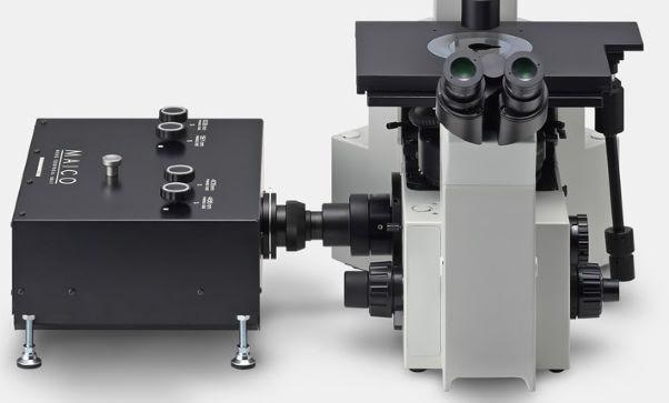
Image Credit: Hamamatsu Photonics Europe
Connecting to a microscope
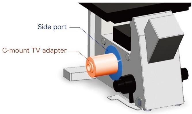
Image Credit: Hamamatsu Photonics Europe
MAICO® is connected to the inverted microscope through a side port and a C-mount TV adapter.
Features
The modular subunit structure makes adding extra channels easy
MAICO® has a one-of-a-kind subunit structure that houses all of the components required for each fluorescence band’s excitation and recognition in a single unit.
When users buy their first unit, for instance, they can choose a single fluorescence band by using the subunit structure. As the research continues, users can add new imaging channels.
Furthermore, MAICO’s sleek design enables subunit installation on-site, minimizing disruption to ongoing research.
MAICO® allows for single-channel observation as well as up to four multi-channel simultaneous excitations and analysis (405 nm, 488 nm, 561 nm, and 638 nm).
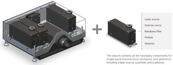
Image Credit: Hamamatsu Photonics Europe
High-sensitivity detectors
The detectors from Hamamatsu Photonics have a long history in fluorescence research. The company was able to decrease the laser power to class 3R with sufficient fluorescence signals by using the world’s most sensitive detector (photomultiplier tube) and utilizing their signal processing knowledge.
This has significant implications for live-cell imaging applications, like long-time-lapse imaging.
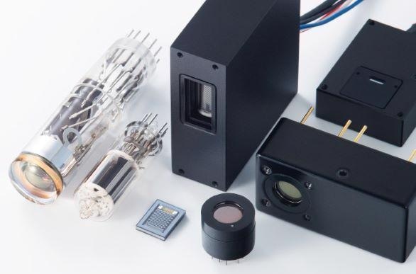
Image Credit: Hamamatsu Photonics Europe
High-speed scanning with MEMS mirror
As a spot scanning device, the 29 kHz resonant type high-speed MEMS mirror was used. The MEMS mirror can scan at up to 76 frames per second, making it ideal for high-speed phenomena like Ca2+ dynamics.
The high-speed resonant scanning system cuts down on laser irradiation time, allowing for minimal phototoxicity, low photobleaching, and high-efficiency monitoring of both living and fixed materials.
When looking for and centering on samples, fast-speed scanning enables comfortable inspection at high resolution with minimal display delay.
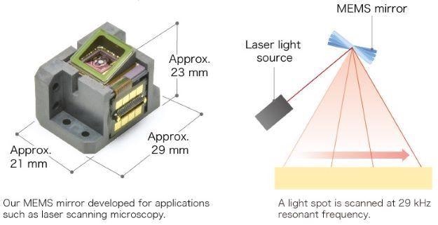
Image Credit: Hamamatsu Photonics Europe
Conventional confocal optics
MAICO® was created to work with traditional confocal optics, which has a long history of acquiring optical sectioning images utilizing a spot scanning device and pinholes.
Only fluorescence emitted from the focus plane of the objective passes through the pinholes to the detector when imaging thick fluorescence samples, whereas fluorescence emitted from further away is prevented.
It recognizes signals that are measurable, repeatable, and dependable. It also captures images with great contrast without the use of image processing techniques like deconvolution.
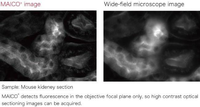
Image Credit: Hamamatsu Photonics Europe
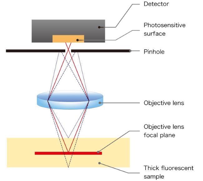
Image Credit: Hamamatsu Photonics Europe
No laser control area required
Hamamatsu was able to lower the laser power to a Class 3R using MAICO’s most sensitive sensors and signal processing expertise. As a result, MAICO can be used in a regular laboratory setting without the need for a laser-controlled area.
Software support
MAICO® can be managed by Hamamatsu’s HCImage software and is consistent with DCAM-API®, a common camera library. Third-party software that is DCAM-API® compliant can also be used to obtain images.
Technology
Enable simultaneous multiband confocal acquisition without bleed-through
Conventional simultaneous multiband observation
Fluorescent dyes emit a wide range of fluorescence wavelengths, and if several fluorescent dyes are utilized, the fluorescence distributions tend to overlap. When multiple dyes are simultaneously excited, an overlapping emission spectrum leaks into adjacent detection channels. This causes an artifact of fluorescence imaging called bleed-through.
The bleed-through cannot be avoided by merely selecting the wavelength using a dichroic mirror or a bandpass filter, and this has been an issue.
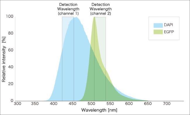
Image Credit: Hamamatsu Photonics Europe
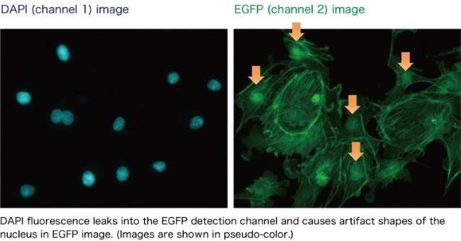
Image Credit: Hamamatsu Photonics Europe
Newly developed: Simultaneous multi-band observation without bleed-through
Researchers can customize the light route in each subunit separately as MAICO® has a subunit structure. This reduces bleed-through to neighboring subunits while allowing for concurrent multiband observation with good detection effectiveness.
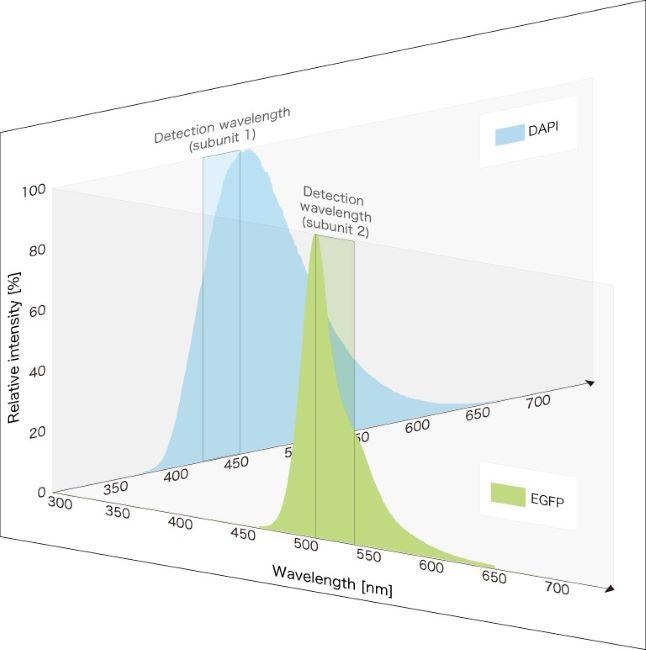
Image Credit: Hamamatsu Photonics Europe
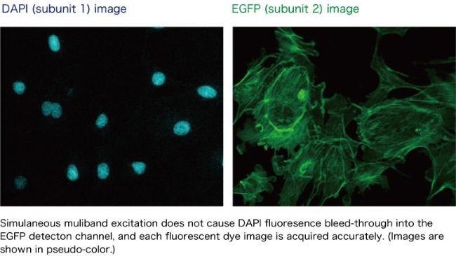
Image Credit: Hamamatsu Photonics Europe