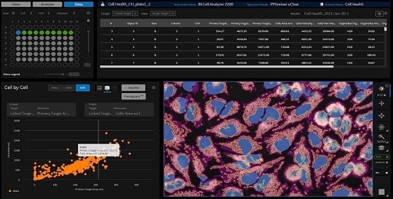
IN Carta® Image Analysis Software from Molecular Devices helps resolve complicated image analysis issues with the help of advanced Artificial Intelligence (AI), which converts images into results that can be interpreted comfortably.
User-friendly workflows help users get answers faster from 2D, 3D, and 4D experiments. Users can design highly customized image analysis methods by integrating the Custom Module Editor application, allowing robust findings to be obtained — even for complicated assays — then quickly display, evaluate, and interact with the analysis results.
Users can focus on their research while letting IN Carta software handle the hard work.
Powerful
Scalable batch processing and guided workflows boost productivity and reduce the time to answer. Experiments can be quickly set up and the analysis of multiple wells can be run in parallel.
Insightful
Machine learning aids users when in comes to harnessing more data and improving precision in the analysis of high-content screening data to enable new discoveries with confidence.
Intuitive
Cutting-edge technology and modern user experience reduce the software learning curve and eliminate barriers to productivity.
IN Carta Image Analysis Software. Image Credit: Molecular Devices UK Ltd.
Features
Deep learning
Enhance the specificity of an image analysis workflow by using the SINAP module. SINAP depends on deep learning-based image analysis, leading to a strong segmentation for virtually any biological structure.
AI-powered data analytics
Users can leverage the power of machine learning without being data scientists. Determine and measure phenotypic changes in a user-friendly workflow. Users can explore their data and disclose insights from complicated datasets. Determine novel and sudden phenotypes with just a few clicks of a mouse.
3D analysis
The 3D application of the Custom Module Editor offers unparalleled flexibility in segmenting complicated biological structures. Image datasets can be acquired in 3D or 4D (timelapse 3D), and customized image analysis routines can be developed within a guided workflow.
Focused worklists
Users can browse the parent directory and populate their worklists with image datasets of interest or use search to locate a dataset of interest.
Customization
Browse and review images from experiments, make image analysis protocols of various complexity, and add on-demand data classification. Envision analytical outcomes using 360 ̊ data connectivity between data tables, images, and charts.
Batch analysis and monitoring
Examine several experiments in batch analysis mode with one or more analysis protocols. Track the status and follow the development of any job that has been submitted.
IN Carta SINAP
SINAP is a module that uses deep learning algorithms to enhance the reliability and precision of high-content screening assays at the segmentation stage, which is the first step in the analysis pipeline. It offers improved object detection compared to conventional image analysis methods.
Deep learning models can be easily tailored within a user-friendly tool to segment any novel biological objects efficiently. Quantitative information removed from segmented objects is highly precise, so errors are not propagated down the analysis pipeline.
With SINAP, segmentation is not a problem
- Accurate: Deep learning has the potential to retain precision throughout hard-to-segment samples such as low signal-to-noise samples, confluent cells, and transmitted light images.
- Flexible: A single workflow can handle a range of applications and imaging modalities.
- Reliable: SINAP models can account for high phenotypic variability.
- Accessible: Instead of requesting a deep learning expert to create a new model and tweak several parameters, the trained model learns to segment from the researcher’s annotations on the image.
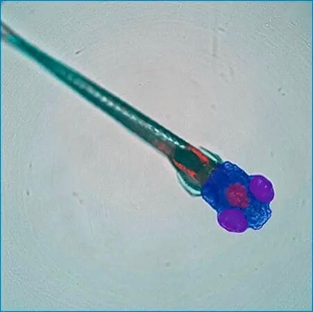
Customized SINAP deep-learning models segment specific regions (whole body, head, eyes, brain) of a zebrafish embryo in transmitted light image. Image Credit: Courtesy of Guo Lab, UCSF.
IN Carta phenoglyphs
IN Carta® Phenoglyphs™ Software Module uses a unique combination of supervised and unsupervised machine learning to measure phenotypical changes. A thorough phenotypic profile has been created and can be used throughout the whole screening procedure with the aid of several hundred different cellular characteristics that can be simultaneously checked.
This multivariate categorization approach provides detailed characterization of object populations, allowing users to resolve minor phenotypic alterations brought on by genetic modification or drug treatment. It can be utilized across many biological targets, including organoids, cells, spheroids, and more.
- Comprehensive: A data-driven approach that starts with unsupervised clustering to identify patterns in the data and highlight subpopulations without having any prior knowledge of potential phenotypes.
- Robust: The enhanced collection of descriptive characteristics is determined by a fragile machine learning technique to avoid overfitting the subsequent classification model.
- Optimized workflow: Classification is achieved by verifying or correcting the predictions of the algorithm until it learns the correct behavior.
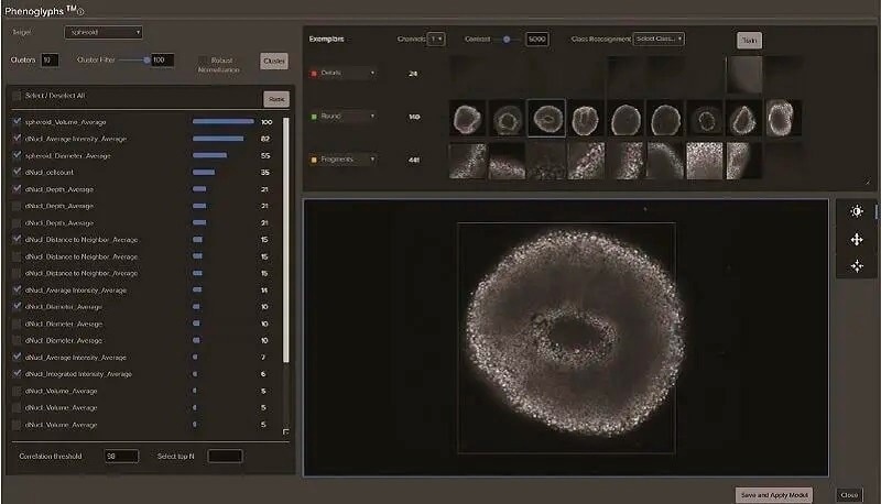
Classification of spheroids formed from HCT116 cells. Spheroids were stained with Hoechst 33342 to visualize nuclei. 3D stacks were collected over time. Image Credit: Molecular Devices UK Ltd.
IN Carta custom module editor 2D and 3D
- Make simple stepwise custom analysis
- True 3D segmentation (3D application only)
- Customize object segmentation and classification
- Only report the measurements that are needed for an assay of interest
- Determine objects that have been localized within specified biological compartments
- Examine live imaging data
- Strong reconstruction of 2D segmentation into volume for 3D assays (3D application only)
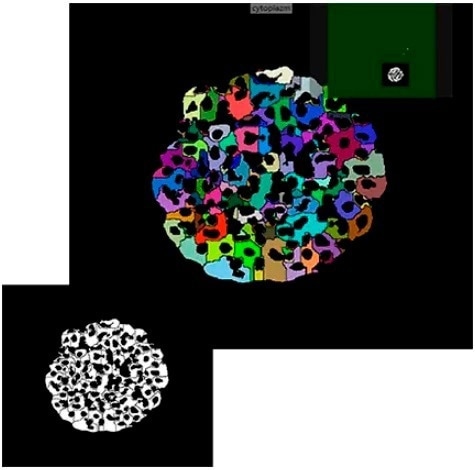
Example segmentation of iPSC-derived hepatocyte spheroids in Custom Module Editor. Image Credit: Molecular Devices UK Ltd.
ImageXpress® confocal HT.ai high-content imaging system
Strong multi-laser light sources, water immersion objectives, a deep tissue penetrating confocal disk module, and modern machine learning analysis software
- Perfect for highly complicated cell-based and 3D assays
- Spinning confocal disk technology for deeper tissue penetration, leading to a sharper image with enhanced resolution
- Seven-channel high-intensity lasers produce brighter images with a greater signal-to-background ratio
- Water immersion objectives provide up to quadruple the signal at lower exposure times for image clarity and higher sensitivity
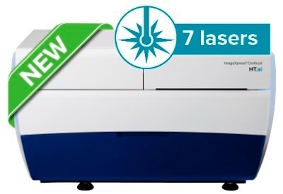
Image Credit: Molecular Devices UK Ltd.
StratoMineR advanced cloud-based analytics
Generate clear, deep data from complex datasets
High-content imaging data can be rapidly transferred into StratoMineR using robust and user-friendly processes. It is then exploited with powerful data mining algorithms to create rich and interactive visualizations.
When utilized with IN Carta Image Analysis Software, it offers strong and quantitative outcomes from complicated biological images and datasets using advanced AI technology. Users can make use of all of their high-content data to find, classify, and examine Phenotypes.
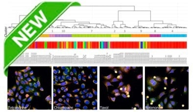
Image Credit: Molecular Devices UK Ltd.