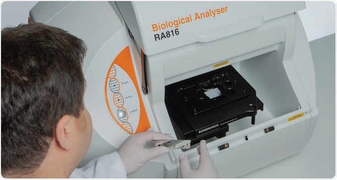
The Renishaw Biological Analyser is a compact, easy to use, benchtop Raman imaging system that redefines tissue and biofluid analysis. It extracts biochemical information contained within the biological samples being analysed.
The system quickly acquires detailed information on the distribution and amount of biochemical species within biological samples including biofluids, tissue biopsies, and tissue sections. It is designed exclusively for the biological community.
The Renishaw Biological Analyer produces outstanding results, quickly and easily. It collects advanced optical and spectroscopic imaging technologies, and the chemical analysis power of Raman spectroscopy (a light scattering technique) in a simple and robust system. In-depth biochemical information is shown from biological samples being studied, from the detection of protein secondary structure alterations due to drug interaction and tissue injury, to the distribution of exogenous and endogenous compounds within tissue.
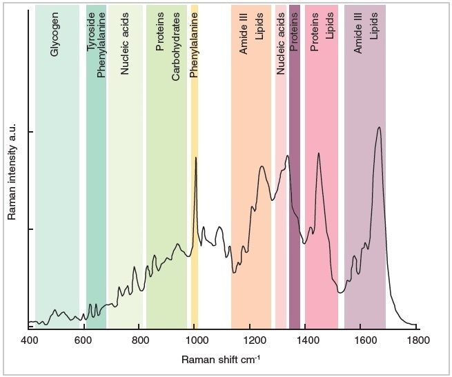
Typical biological tissue Raman spectrum demonstrating the wealth of information obtained from a single measurement
Raman spectroscopy provides many benefits for the study of biological materials:
- No labeling or sample staining needed
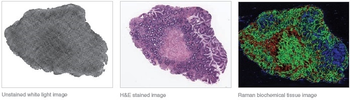
- The high-resolution Raman images generated give biomolecular information but also retain the spatial morphology
- Non-invasive optical technique (retains sample integrity)
- Measures multiple molecular constituents in biological samples at once (saves cost and time)
- High specificity: assists in discovery and validation of early onset disease markers
The Renishaw Biological Analyser has a range of features making it the ideal tool for clinical research:
High Performance for Demanding Applications
- Post measurement check option to validate performance stability over the measurement period
- LiveTrack™ technology to track sample surface and retain focus
- Streamline™ technology for high speed data collection and image generation, without causing laser sample damage
- Stable and repeatable, with integrated performance qualification (PQ) and alignment
- High throughput with high signal to noise spectral data and optimised substrates
Easy to Use
- Reflection or transmission illumination for best image contrast
- Image tiling for large area coverage
- Easy to use software with sample concept and workflow: making Raman spectroscopy accessible to all users, without compromising performance
- Macro and high magnification modes, with digital zooming for a range of sample sizes
Dedicated Data Acquisition and Analysis Software
- Enable configuration of multiple measurements for unattended operation
- Optimal system and sample workflow: enables robust, repeatable and reproducible data to be collected and analysed
- Experiment templates for different biological sample types to simplify operation
- Model building software for clinical data classification: provides the ability to build and validate pathology and disease models
- Validate the model with new samples enabling the stratification and classification of unknown tissue and biofluid samples
Compact, Robust and Transportable
- Robust system with no user alignment
- System can be easily moved between lab and clinic with inbuilt performance checking to ensure accurate operation
- Small footprint - ideal for a space conscious laboratory environment
The Ideal Toolkit for Translating Raman to the Clinic
- Bulk tissue biopsy sample holder eliminates the need for tissue sample preparation (e.g. cryosectioning or microtoming)
- Performance standards support data transferability of classification models on additional Renishaw Biological Analyser systems
- Low-cost, ultra-low background disposable mirror slides for enhanced reflection, to increase Raman signal and improve white light contrast
- Specially designed bio-sample holders and inserts for bio-fluids/liquids and tissue sections
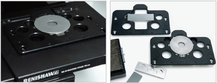
A range of specially designed sample holders
Powerful Software – Data Acquisition
The Renishaw Biological Analyser’s software gives a structure for managing every step of the measurement process, using predefined protocols and experiment setups. It is completely computer-controlled. The system can acquire data unattended. Its queuing capability enables you to configure measurements and leave the instrument to run them; you can analyse multiple samples on a slide without the need for user intervention. The unique macro image provides a comprehensive overview of all subsequent work and enables easy sample navigation and visualisation.
Building and Validating a Classification Model
The system contains a classification software package that enables users to:
- Minimise instrument and sample quality variations within and between the instruments and sites
- Data process spectra datasets
- Test and validate classification models with new independent sample data
- Build, test and validate pathology and disease classification models (using PCA-LDA)
These tools establish early onset disease markers, assist in the discrimination of cancer stages with high sensitivity and specificity, and identify biochemical adjustments that are associated with cancer formation and progression.
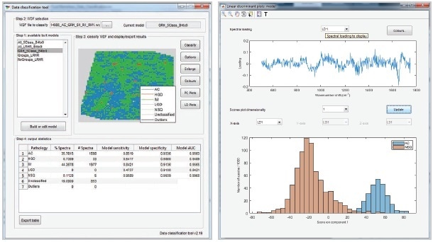
Renishaw data classification software
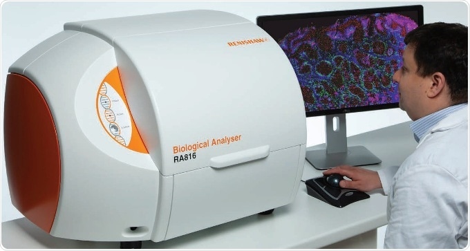
The Renishaw Biological Analyser
Medical research using Raman Spectroscopy
Example Workflows for Routine Operation
Raman Tissue Imaging and correlating results with stained/labelled slides
- Deposit the stained/ labeled slide in its dedicated holder and select from the list of possible bio-sample types
- Using reflection or transmission white light imaging (macro view or high-resolution tile), the system automatically digitises the slide
- Insert and digitise an adjacent unstained tissue section
- Based on areas of interest from the stained image, select the whole sections or regions for Raman analysis on the unstained image
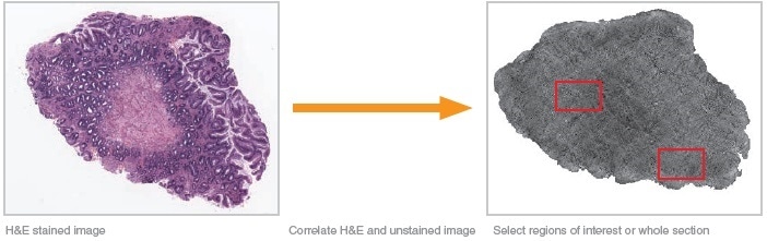
- The system then:
- Scans the selected areas or tissue section (using LiveTrack focus-tracking technology to maintain highest signal quality and keep in focus, if necessary)
- Creates biochemical images that show morphological and chemical information about the tissue sample
The Renishaw Biological Analyser
Raman Spectral Data Collection for Disease/Pathology Classification
- Place your tissue or biofluid samples on a dedicated sample holder
- The system generates a macro image of the whole slide automatically (providing an overview of the array of samples)
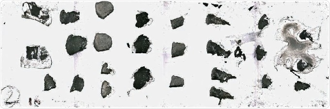
Macro image of slide
- If necessary, generate a high-resolution tiled image of the individual tissue sections
- Identify the points or area to analyse and start analysis utilising predefined measurement settings
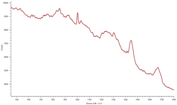
Mean of example spectral dataset (taken from data measured at 4s time exposure;1 accumulation; 100% laser power)
- Using the inbuilt data classification tool, perform multivariate analysis to reveal biochemical information about the tissue samples, and generate classification performances of the disease and pathology models
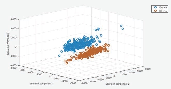
Example Multivariate analysis (PCA) of IDH genomic mutation status of brain glioma
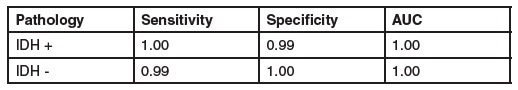
IDH genomic mutation status classification performance using the Renishaw data classification software*1
*1 Livermore et al. 2018 manuscript in preparation.
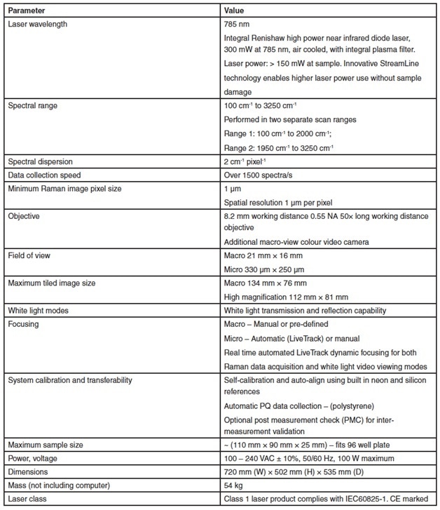
Specifications - The Renishaw Biological Analyser RA816
The Renishaw Biological Analyser is designed for research use only (RUO) and is not for use in diagnostic procedures.