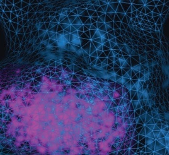
The Vutara VXL comprehensive biological workstation from Bruker allows core facilities and separate investigators to seamlessly integrate super-resolution microscopy into their research.
The system utilizes single-molecule localization microscopy (SMLM) technology for the highest resolution of all super-resolution techniques.
When integrated with Bruker’s microfluidics unit, Vutara VXL enables multiplexed imaging for transcriptomics, targeted spatial genomics, and proteomics research. Using sophisticated SRX software, scientists can convert complex data into useful information to explore new frontiers in disciplines varying from neuroscience and cell biology to virology.
Image Credit: Bruker Nano Surfaces and Metrology
The Vutara VXL advantage:
- The SMLM method offers the highest resolution, down to 20 nm laterally
- Innovative top-hat illumination allows even and quantitative data collection
- Proprietary Biplane™ technology helps obtain the deepest 3D imaging, with depths of up to 50 μm
Leading super-resolution technology
Approach: Single-molecule localization
For more than 10 years, Bruker has been leading super-resolution technology, providing the highest quality and most advanced microscopy systems on the market.
Vutara VXL uses single-molecule localization (SML), which offers the highest resolution compared to other super-resolution approaches, down to 20 nm laterally.
For the optical resolution limit with SMLM to be overcome, just a limited number of chromophores are triggered at a time so that their point-spread functions (PSF) do not intersect.
The microscope detects the presence of these chromophores, identifies their location, and switches them off. Another subset of chromophores is triggered, localized, and deactivated. This cycle has been repeated several times to create a high-resolution localization map.
It is possible to achieve stochastic excitation of molecules using organic fluorescent dyes or fluorescently labeled proteins. Vutara VXL is entirely consistent with standard and developing SMLM methods, including:
- Photoactivated localization microscopy (PALM) is available
- Stochastic optical reconstruction microscopy (STORM) and direct-STORM (dSTORM)
- Points accumulation can be done for imaging in DNA-PAINT and nanoscale topography (PAINT)
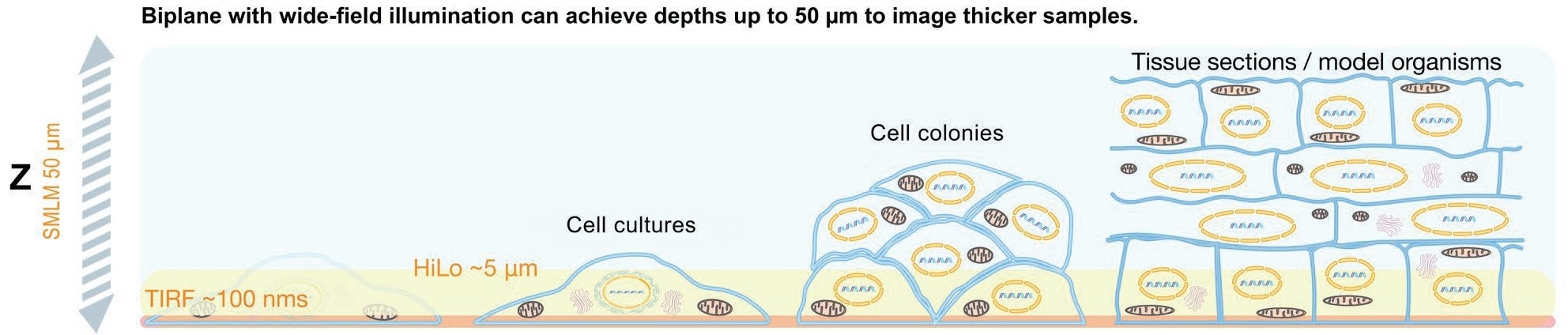
Image Credit: Bruker Nano Surfaces and Metrology
Innovation for unlimited potential
Vutara VXL has been developed in partnership with leaders in industry and academia and pushes the boundaries of standard super-resolution capabilities. Proprietary biplane technology, integrated with a spatial filter in the emission light path, enables users to obtain 3D data up to 50 μm deep into the sample.
The system enables scientists to execute a Z series efficiently and automatically localizes and reconstructs the complete volume. Top-hat illumination leads to even data collection throughout the imaging region, allowing trustworthy quantitative data analysis. Vutara VXL was designed to be an all-embracing biological workstation, customized to the requirements of microscopy experiments from start to finish.
The imaging ecosystem—such as the Vutara VXL microscope, the microfluidics unit, and the SRX software—aids smooth acquisition and analysis of rich, quantitative data.
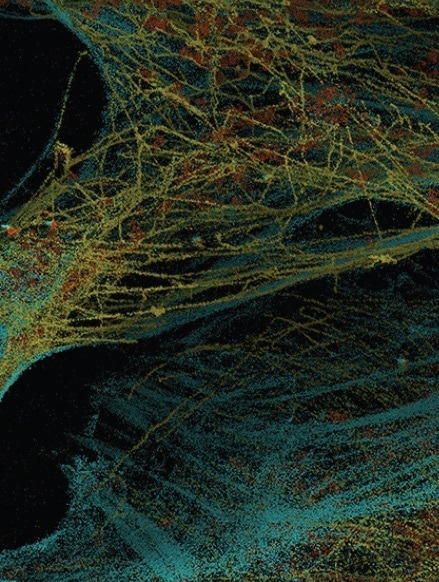
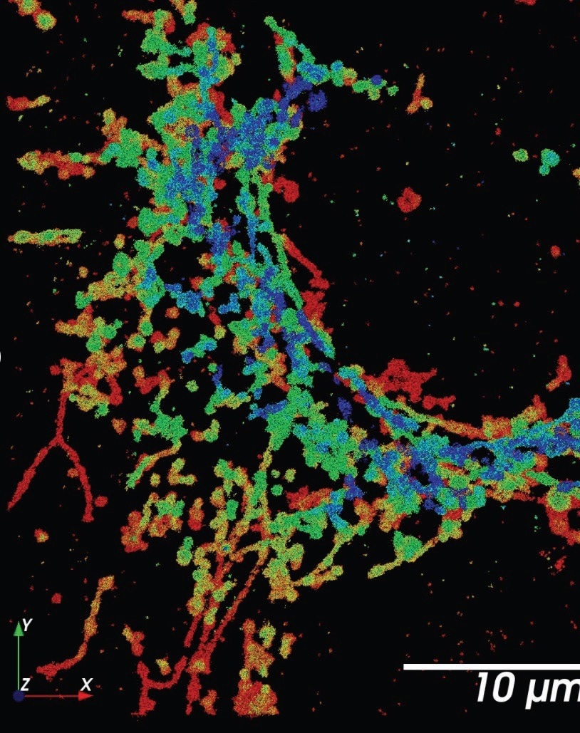
Actin-magenta, microtubule-yellow, mitochondria-orange. Image Credit: Bruker Nano Surfaces and Metrology
Enabling advanced research across the life sciences
Vutara VXL allows research on RNA, DNA, and proteins, from macromolecular complexes and super-structures to chromosomal substructures and chromatin structure to studying functional relationships in genomes and several subcellular organelles.
This novel system aids sophisticated spatial biology research in extracellular matrix structures, virology, neuroscience, extracellular vesicles, and live-cell imaging. When integrated with Bruker’s special microscope fluidics unit, Vutara VXL assists multiplexed imaging for targeted, transcriptomics, submicrometer multiomics in genomics, and proteomics research.
Neuroscience
Imaging the brain in nanoscale detail far above the optical diffraction limit sets the stage in neuroscience research. One of the most fascinating components of the brain is the synapse—known to be the junction necessary for the transmission of the brain’s signals.
Super-resolution microscopy is needed to envision such structures approximately 20 to 40 nm wide. The SML method is perfect for the study of neuronal structures and processes such as:
- Neural development
- Synaptic formation
- Synaptic transmission
- Neural circuit assembly
- Synaptic proteins
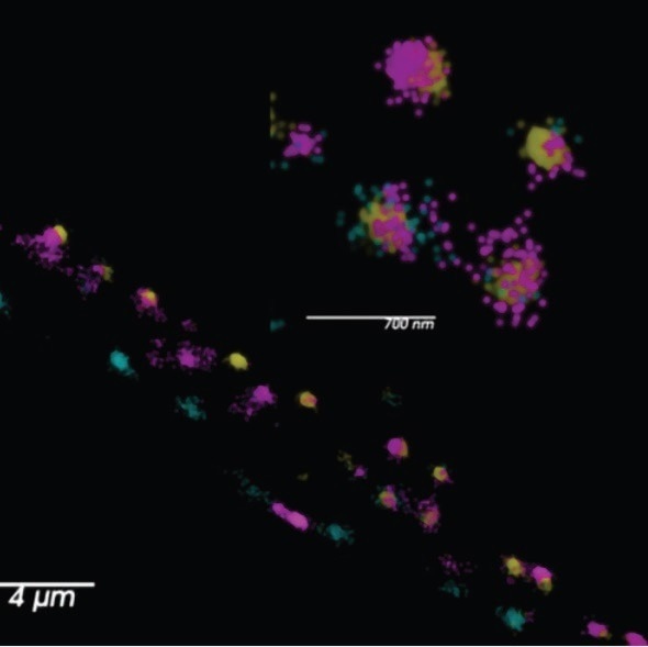
The synaptic labeling of C. elegans. Three markers are present in this image: yellow (Skylan-s Active zone marker), magenta (HaloTag::JF646 Calcium Channel 1), and cyan (SNAPf::JF549 Calcium Channel 2). Image Credit: Dr. Sean Merrill and Dr. Erik Jorgensen, University of Utah
Genomics
Envisioning genomic organization and structure on a subchromosomal level is essential for comprehending relationships between genes, their control, and their surroundings. SMLM can image three-dimensional chromosome structures at the single cell and subcellular levels, making it difficult for genomics applications such as:
- Functional organization of genome within the nucleus
- Multi-location capture of multiple nuclei
- Chromatin tracing
Chromosome 19 topologically associating domain (TAD, in magenta) within a compartment (in blue). Image Credit: Ting Wu Lab, Harvard University.
Virology
The Vutara SMLM platform has been a vital instrument for comprehending viral particles. Typically, viral particles are much smaller than the optical diffraction limit, making SMLM the ideally suited fluorescence method for resolving virus particle structural details or identifying the localization of virus components with the cellular machinery. A few main applications for Vutara VXL in virology research include:
- Viral particle structure
- Virus pathology
- Virus-host interactions
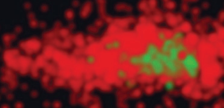
Mitochondria stained for TOM20 with Alexa 647, imaged with SMLM (dSTORM). Image Credit: Bruker Nano Surfaces and Metrology
Cell biology
Several fascinating cellular structures are smaller than the optical diffraction limit of approximately 200 to 300 nm and hence need super-resolution microscopy for imaging specifically labeled systems at this scale.
These consist of the substructures of most organelles and all macromolecular machines, channels, and receptors. Research applications in cell biology that could be obtained with Vutara VXL include:
- Molecular quantification
- Colocalization of proteins
- Molecular distribution
- Particle tracking
Vesicular stomatitis viral particle. Red – VSV-G protein tagged with Alexa Fluor 647. Image Credit: Bruker Nano Surfaces and Metrology
Transforming Voxels into information
With SRX software and its quantitative localization microscopy analysis suite, Vutara VXL delivers visual and quantitative biological sample information. Vutara can generate stunning 3D images by localizing individual molecules while providing in-depth quantitative analysis tools. Measurements available within the SRX software include:
Spatial Distribution—Offers a range of tools for examining spatial distribution relationships of particles, such as pair correlation, Ripley’s K, and nearest neighbors
Cluster Analysis—Cluster sizes, counts clusters, and cluster densities Colocalization—offers statistical measures on relationships occurring between particles of two various labels.
Resolution Analysis—Measures resolution for images that have been derived from localized data
Live-Cell Analysis—It is possible to monitor clusters in live-cell experiments with angular displacement analyses and mean squared displacement.
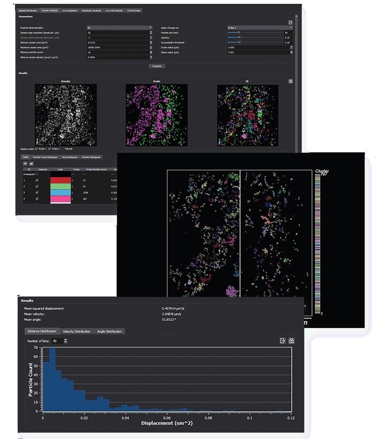
The workflow-oriented SRX software includes powerful statistical analysis tools. Image Credit: Bruker Nano Surfaces and Metrology
Multiplexing for unlimited imaging possibilities
The microfluidics unit has been developed to integrate sequential labeling into localization microscopy protocols with the Vutara VXL microscope.
The unit is controlled by SRX software, enabling the adaptable configuration of fluidics sequences to run jointly with imaging cycles. This allows users to label an endless number of probes and image them.
With a microfluidics unit, Vutara VXL could be utilized to fulfill any localization microscopy application that needs numerous targets to be imaged. A total of 15 probe vials have been offered and could be exchanged during experiments, assisting a limitless number of targets.
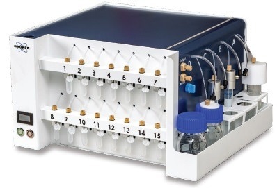
Image Credit: Bruker Nano Surfaces and Metrology
Listed below are the primary localization microscopy applications with the microfluidics unit:
- OligoSTORM
- DNA-PAINT
- Multiplexed
- RefreshSTORM
Specifications
Source: Bruker Nano Surfaces and Metrology
| . |
. |
| Imaging Modalities |
SMLM with high Z-resolution for STORM, PALM, PAINT, and related super-resolution applications;
Localization microscopy with large FOV, optimized for super-resolution multiplexed genomics applications;
Epi-fluorescence microscopy with large FOV;
Transmitted light microscopy with large FOV |
Excitation lasers
(nominal laser power at diode) |
405 nm, 120 mW
488 nm, 2000 mW (optional)
555 nm, 2000 mW
638 nm, 1200 mW
750 nm, 1500 mW (optional) |
| Flat Illumination |
Flat excitation profile guaranteed by top-hat illumination from a square fiber |
| Multi-Color Acquisition |
≤5 colors sequential |
| Multi-Plane Imaging |
Simultaneous imaging of two focal planes allows 1 μm depth discrimination (larger Z range possible in Z-stack mode) |
| Camera |
Orca Flash 4.0 v3 sCMOS camera; Orca Fusion BT sCMOS camera (optional) |
| Objective |
60x magnification; 1.3 NA; Silicon oil immersion; 0.3 mm working distance;
#1.5H cover glass (0.170 ±0.005 mm) |
| Field of View (FOV) |
200 x 200 μm for multiplexed localization microscopy and widefield imaging; 50 x 50 μm for SMLM with switching (STORM, PALM, PAINT) and 3D localization (Biplane detection); Larger FOV with tile scanning |
| SMLM Resolution |
20 nm laterally (XY); 50 nm axially (Z) with Biplane |
| Imaging Depth |
>30 μm (dependent on sample) |
| XY-Stage |
Easy access to sample; 100 x 50 mm travel range; Optical encoder with 1 nm resolution |
| Z-Focus |
Course focus to localize sample; Fine focus for fast Z-stack acquisition |
| Drift Correction |
Active focus drift correction during data acquisition; Focus drift <30 nm over 10 minutes;
XY drift correction post-acquisition |
| Workflow-Oriented Software |
Data acquisition; 3D localization based on measured PSF; Particle tracking; Statistical data analysis tools; Visualization |
| Options |
Integrated microfluidics for multiplexed experiments; Live-cell incubation with humidity, CO2, and temperature control; Network attached storage |