Oxygen is a vital element for human life, and lack of it leads to death in just a few minutes. A condition where the respiratory system fails in one or both of its gas exchange functions (oxygenation and elimination of carbon dioxide from mixed venous blood) is known as respiratory failure. One of the main indicators of such failure is hypoxemia – a significant drop in blood oxygenation.
The whole process of oxygenation is, akin to other physiologic processes, dynamic. Similar to other organ systems, the human lung can respond to injury in only a limited number of ways. Consequently, five main pulmonary mechanisms result in arterial hypoxemia: ventilation-perfusion mismatch, diffusion impairment, alveolar hypoventilation, right to left shunt, and diffusion-perfusion abnormality.

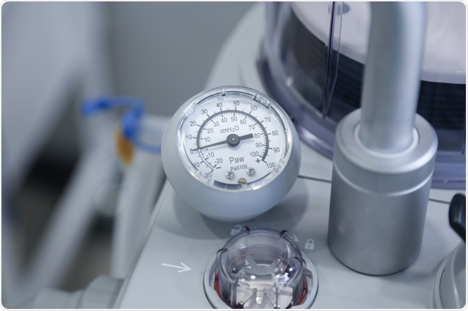
Image Credit: photos_adil / Shutterstock
Difference between hypoxemia and hypoxia
Although the terms hypoxemia and hypoxia are often used interchangeably, they differ from each other. Hypoxemia can be defined as a condition where arterial oxygen tension or partial pressure of oxygen (PaO2) is below normal (normal value is between 80 and 100 mmHg). On the other hand, hypoxia is defined as reducing oxygen supply at the tissue level, which is not measured directly by a laboratory value.
Generally, the presence of hypoxemia suggests hypoxia. However, hypoxia may not be present in hypoxia patients if the patient compensates for a low PaO2 by increasing oxygen delivery. This is typically achieved by increasing cardiac output (by raising the heart rate) or decreasing tissue oxygen consumption.
Conversely, patients who do not show signs of hypoxemia can be hypoxic if oxygen delivery to tissues is hampered or the tissues cannot adequately use oxygen. Cyanide poisoning is a classic example of the latter because cyanide interferes with aerobic metabolism in the tissues. Nevertheless, hypoxemia is definitely the most common cause of tissue hypoxia, and the correct diagnosis should be made because it is readily treatable.
Clinical assessment of hypoxemia
As assessing proper oxygenation is often difficult, a good history is an important first step. Patients most susceptible to hypoxemia have a history of respiratory and cardiac illness and smoking. History taking should lead to measurement of arterial oxygen tension (PaO2) and percent saturation of hemoglobin with oxygen (SaO2).
In normal conditions, breathing accounts for a minimal fraction of the patient’s total oxygen consumption. Most hypoxemic patients complain of shortness of breath and dyspnea. Thus the work of breathing is significant enough to be noticed by the affected patient. Also, non-specific changes in mental status are often seen in severe hypoxemia.
Vital sign abnormalities in hypoxemia include tachycardia (heart rate faster than 100/min) and tachypnea (a respiratory rate higher than 24/min). Still, patients with a limited cardiac reserve may not manifest with tachycardia. In severe cases of hypoxemia, even bradycardia (i.e. abnormally slow heart rate) may ensue due to vagal stimulation.
Patients with central nervous system disease can even have a normal breathing pattern. Hence the absence of the aforementioned tachycardia and tachypnea should never be considered as a certain sign that the patient is not hypoxemic. Oxygen content represents the minimal laboratory information needed to assess proper oxygenation, which is a value that takes into account the amount of hemoglobin available to carry the oxygen throughout the body.
Sources
- http://erj.ersjournals.com/content/22/47_suppl/3s.long
- http://onlinelibrary.wiley.com/doi/10.1002/lt.500020412/pdf
- https://www.sallyosborne.com/
- http://onlinelibrary.wiley.com/doi/10.1111/resp.12204/full
- Samuel J, Franklin C. Hypoxemia and Hypoxia. In: Myers JA, Millikan KW, Saclarides TJ. Common Surgical Diseases – An Algorithmic Approach to Problem Solving. 2nd edition, Springer New York, 2008; pp. 391-394.
Further Reading
Last Updated: Feb 17, 2023