Microfluidic Sanger sequencing is the implementation of the Sanger method of DNA sequencing on a chip; therefore, this allows for the manipulation of fluids at the submicron size. Requiring much smaller sample volumes, microfluidic Sanger sequencing automates the reaction, separation and detection processes. The smaller scaling provides the advantage of reducing diffusion times and the surface-area-to-volume ratio means that the transference of heat is enhanced as size is reduced.
Credit: ktdesign/Shutterstock.com
Furthermore, microfluidic systems at the micro- and nano-scale more accurately reflect the cellular environment, as physicochemical transport processes of most biological systems function at the same scale. As chips can be manufactured cheaply in large numbers, the benefits of this technology are being applied to laboratories in biomolecular fields such as genetic analysis.
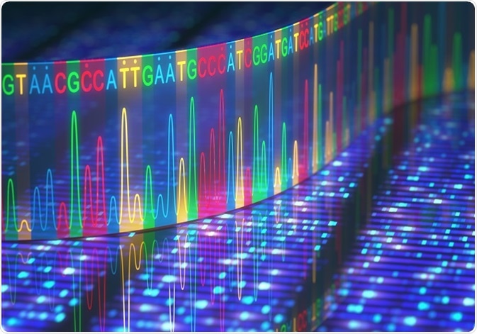
DNA sequencing via microfluidic Sanger sequencing
Though there are other high through-put methods of sequencing DNA, microfluidic Sanger sequencing is a robust method, with a low error rate and long read lengths (700-800 base pairs). This makes the method particularly suitable for sequencing large new genomes or genome segments that are highly rearranged. Sanger sequencing works by incorporating a fluorescently labeled deoxynucleotide to the terminal end of a DNA fragment. A sequence can then be formed with the DNA fragments placed in order by the base-specific fluorescent label.
The microfluidic chip incorporates all the important processes of Sanger sequencing, including thermal cycling and sample purification before separation by electrophoresis. Fully integrated microfluidic devices allow for efficient connections between laboratory processes, minimizing loss and dilution of samples. Further developments of the process on-chip have formed the ability to sequence by denaturation. The fluorescently labeled Sanger fragments, which are end base specific, denature sequentially by heating. As the temperature of denaturation correlates with the number of bases, the decrease in fluorescent signal provides the base sequence.
Fully integrated microfluidic devices and genome sequencing
There is an expanding demand for medical and personal applications of genome sequencing, with the quest for personalized medicine ongoing. Genetic variants found in an individual genome have the ability to be used as markers for:
- Diagnosis.
- Prognosis.
- Disorder prevention.
- Treatment targets.
Future advancements in this field will require the fundamental step of DNA sequencing. Whilst other DNA sequencing methods have a higher throughput, microfluidic Sanger sequencing achieves rapid and low-cost DNA sequencing from single copies of DNA template, which makes the technique particularly suited to sequencing large genomes from single cells.
The Microbead Integrated DNA Sequencing (MINDS) bioprocessor integrates the technologies required for amplifying a single template and then performing Sanger sequencing. Single-copy DNA template is amplified with primer functionalized microbeads in a nanoliter solution. The clonal beards produced are then introduced to the MINDS bioprocessor which performs integrated Sanger sequencing. The nanoliter-volume integrated sequencing system has been shown to save cost, time and space in comparison to conventional whole genome Sanger sequencing. The microfluidic device provides the potential for engineering a workable system that can be used for medium through-put personal sequencing.
Microfluidic devices and forensic DNA profiling
DNA sequencing is also required for the examination of short tandem repeats (STRs). STRs are short sequences of genetic variation called polymorphisms that can vary from person to person. The most variable STRs are useful for human identification purposes and are routinely used in forensic cases. Due to this, microfluidic devices have the ability to hasten forensic DNA profiling from crime scenes. The multiple laboratory techniques integrated into a chip provide a fast analysis time. The chips hold the sample in a closed microfluidic environment reducing the risk of cross-contamination. Microfluidic devices are both portable and frequently designed for single use, meaning they provide the possibility of analysis at the crime scene with reduced risk of contamination. However, most input types for microfluidic devices are either lysate or already purified genomic material, requiring pre-preparation. Some commercial devices have been developed that use buccal swabs as input, but will require standardizing for evidence validation.
Sources
- Yeo, L.Y. et al. 2011. Microfluidic devices for bioapplications. Small, 3, pp. 12-48. http://onlinelibrary.wiley.com/doi/10.1002/smll.201000946/abstract
- Offit, K. 2011. Personalized medicine: new genomics, old lessons. Human Genetics, 130, pp. 3-14. https://www.ncbi.nlm.nih.gov/pmc/articles/PMC3128266/
- Bruijns, B. et al. 2016. Microfluidic devices for forensic DNA analysis, Biosensors, 6, e41. http://www.mdpi.com/2079-6374/6/3/41
- Liu, P. & Mathies, R.A. 2009. Integrated microfluidic systems for high-performance genetic analysis, Trends in Biotechnology, 27, pp. 572-581. http://www.sciencedirect.com/science/article/pii/S0167779909001206
Further Reading
Last Updated: Feb 26, 2019
 Standard DNA tests miss most cases of NUT carcinoma