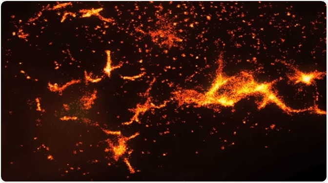
Photoactivated Localization Microscopy (PALM) is a form of super resolution fluorescence microscopy (SR) which enables highly resolved imaging to be produced through the selective targeting of fluorescent markers within a specimen.

Super-resolution microscopy
The technique differed from more conventional microscopy techniques that were available at the time of its development as it was able to produce imaging beyond the diffraction limit (the point at which molecules 200 nm or less in size can be distinguished from one another).
The Super-Resolution Revolution
Through exposure to ultraviolet light, proteins within a specimen can be excited so that they are photoactivated. This enables the position of the photoactive proteins to be localized. Having been exposed to ultraviolet light for a prolonged period, these photoactivated proteins will be bleached and no longer fluoresce. The process can be repeated until all the proteins within a specimen are localized.
Like other SR techniques, PALM has various applications and has had a massive impact on biomedical research.
An introduction to super-resolution microscopy of living cells
PALM in HIV Studies
Human immunodeficiency virus is a retrovirus which attacks T cells. It is transmitted through certain bodily fluids. The functional T cells are diminished as the condition progresses, leaving individuals who live with HIV susceptible to various forms of infection and some types of cancer. The virus remains a global health issue with 1 million people losing their lives in 2016 to HIV-related conditions.
HIV can be categorize into two types, HIV-1 and HIV-2. This classification is based on the viral antigens and characteristics belonging to each type. Globally more than 30 million people live with HIV-1. This type of HIV evolved from an immunodeficiency virus found in Central African chimpanzees known as SIVcpz.
Gag is a polyprotein responsible for a variety of key functions in the assembly process of HIV-1. It enables the assembly of immature particles at the plasma membrane of cells; recruits other viruses to the budding sites (the areas where viruses can exit the host cell) and causes virion maturation to occur. Using PALM, Gag clusters on cells have been observed. These clusters categorized into two separate groups; large, immobile clusters responsible for viral assembly and mobile, individual Gag molecules. Approximately 40% of clusters were represented by smaller and/or individual Gag clusters. This suggests that a transition from a small cluster to a larger one at a viral assembly site may be a rate limiting step in HIV particle formation.
Structural Architecture of Mitochondria
Mitochondria are crucial for the generation of energy in eukaryotic cells. This has been the subject of many studies using SR techniques due to the enhanced resolution provided by these forms of microscopy in comparison to older techniques.
The mitochondrial structure was first observed using PALM. The cytochrome c oxidase system located in the matrix of mitochondria was studied, allowing researchers to better understand its spatial arrangement. This paved the way for further studies using SR to scrutinize other aspects of mitochondrial architecture which had previously been unclear.
Understanding Focal Adhesion
Focal adhesion can be described as the interaction or adhesive contact between the extracellular matrix and the cell. This is facilitated by integrins and scaffold proteins. Using a mode of PALM known as interferometric PALM (iPALM), focal adhesions were able to be localized and imaged. When compared to biochemical data, the imaging produced by iPALM was found to be consistent. It also provided new information regarding the spatial organization of focal adhesions.
PALM has had a profound impact on biomedical research and has proven to have varied applications. Despite this, the technique is still comparatively new, with the prospect of being adapted and changed to better meet the needs of those who require super-resolved imaging as part of their research.
Further Reading
Last Updated: Aug 23, 2018