By Anusha Krishnan, PhD
Single Molecule Localization Microscopy (SMLM) is a class of super-resolution microscopy techniques used to obtain optical images at very high spatial resolutions ranging between 2–25 nanometers.
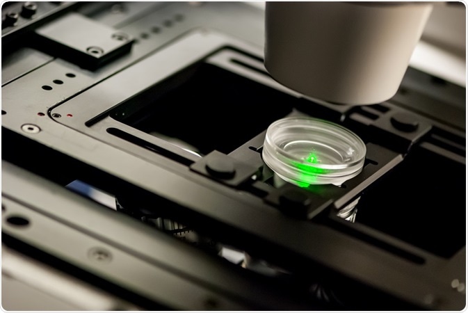 Credit: Micha Weber/Shutterstock.com
In the last decade, rapid advances in SMLM methodologies have allowed scientists to observe intracellular processes - such as the movement of myosin V across actin filaments, single mRNA transcription events, and the dynamics of viral endocytosis - that were once thought to be impossible to visualize.
SMLM history
The super-resolution microscopy was discovered towards the end of the 19th century. Before this, resolving two microscopic objects situated closer to each other than half the wavelength of light was commonly thought to be impossible.
This is the “Abbe limit” or “diffraction limit” for microscopy due to which scientists could not clearly visualize structures smaller than 200 nm (half the wavelength of far blue light, which has a wavelength of 400nm).
In 2014, William Moerner, Stefan Hell, and Eric Betzig were awarded the Nobel Prize in Chemistry for developing radical types of fluorescence-based microscopies that could provide resolution far beyond those imposed by Abbe’s limit.
While Stefan Hell developed STED (Stimulated Emission Depletion) fluorescence microscopy, Moerner’s and Betzig’s work independently laid the foundations of single molecule microscopy.
Principles of SMLM
Super-resolution microscopy is based on the knowledge that the position of a single fluorophore can be located much more precisely than the ~200 nm range dictated by the diffraction limit.
The resolving power of any microscope is determined by the wavelength of light used and the numerical aperture or light collecting ability of the objective lens. Due to these two factors, any point source of light that is smaller than the diffraction limit will appear to be blurry and larger than it really is.
The PSF (Point Spread Function) of an instrument is a characteristic that describes this “blur effect”, or how an object smaller than the diffraction limit appears under certain imaging conditions. PSF is characterized by measuring the apparent width of a fluorescent object at a point where its intensity is 50% of the maximum, called the FWHM (Full Width at Half Maximum).
Using FWHM, the position of a single fluorescent molecule can be obtained with a precision as low as 1 nm. This precision is mainly limited by the number of photons collected at each point and is equivalent to the FWHM divided by the square root of the number of photons collected.
Although visualizing sparse single fluorophores is an excellent method to employ when studying the dynamics of specific target molecules, it is not very useful in studying complex biological systems that are highly crowded and dense. The key to obtaining super-resolution in such systems is the temporal separation of actively fluorescing molecules, which forms the basis of SMLM.
All SMLM techniques are grounded on the principle of temporally modulating the emission of a small set of fluorophores. A low-intensity laser beam is used to stochastically activate, localize, and reversibly or irreversibly bleach a few fluorophores at any given point of time; repeating this process many thousands of times provides highly resolved detail-rich images.
Types of SMLM
SMLM techniques mainly differ in the types of fluorophores used, and the methods used to temporally control the emission patterns of these fluorophores.
PALM (PhotoActivated Localization Microscopy), FPALM (Fluorescence PhotoActivated Localization Microscopy), and STORM (STochastic Optical Reconstruction Microscopy)
All three techniques use the same methodology of activating fluorophores with a high-intensity laser beam, following which a low-intensity laser is used to stochastically excite fluorescence in a sparse set of fluorophores. However, several differences between these techniques exist.
PALM and FPALM originally used proteins such as PA-GFP (PhotoActivatable Green Fluorescent Protein) as fluorophores, whereas STORM used the synthetic carbocyanine dyes Cy3 and Cy5.
Furthermore, in PALM and FPALM, the fluorophore is generally expressed by the cell as a fusion protein tagged to the protein of interest, whereas, endogenous cellular elements are immunolabelled using antibodies tagged with fluorophores in STORM. Nevertheless, it is possible to use synthetic dyes in PALM experiments and fluorescent proteins for STORM experiments.
In addition to these differences, the life of a fluorophore is generally limited due to the photoactivation and photobleaching processes in PALM and FPALM.
In STORM, however, the phenomenon of photoblinking (where the fluorophore switches randomly between the OFF or dark state to the ON or emitting state under continuous excitation) is utilized for temporal separation of fluorescence activation.
FPALM differs from PALM and STORM in the imaging technique used—FPALM uses traditional confocal fluorescence microscopy, whereas PALM and STORM utilize TIRFM (Total Internal Reflection Fluorescence Microscopy).
In TIRFM, fluorophores bound to the surface of cells/tissues in contact with a glass coverslip or the wall of the container are selectively excited; this reduces the number of excited fluorophores in the sample, making the technique ideal for single molecule detection.
COLD (Cryogenic Optical Localization in 3 Dimensions)
COLD, developed in 2016 takes fluorescence microscopy to its fundamental limit as dictated by the size of the fluorophore. COLD allows the sub-molecular localization of multiple fluorescent sites within a single protein molecule, providing resolution at the Angstrom level.
The technique uses low temperatures to slow down photochemical reactions in fluorophores; this allows the emission of higher numbers of photons before photobleaching, hence enhancing localization precision. The 2-dimensional images of fluorophores can be used to reconstruct 3-dimensional configurations using algorithms borrowed from electron microscopy.
Sources:
- Betzig, E., Patterson, G.H., Sougrat, R., Lindwasser, O.W., Olenych, S., Bonifacino, J.S., Davidson, M.W., Lippincott-Schwartz, J. and Hess, H.F., 2006. Imaging intracellular fluorescent proteins at nanometer resolution. Science, 313(5793), pp.1642-1645.
- Hess, S.T., Girirajan, T.P. and Mason, M.D., 2006. Ultra-high resolution imaging by fluorescence photoactivation localization microscopy. Biophysical journal, 91(11), pp.4258-4272.
- Rust, M.J., Bates, M. and Zhuang, X., 2006. Sub-diffraction-limit imaging by stochastic optical reconstruction microscopy (STORM). Nature methods, 3(10), p.793.
- Dickson, R. M., Cubitt, A. B., Tsien R, Y. and Moerner, W. E., 1997. On/off blinking and switching behavior of single molecules of green fluorescent protein. Nature 388:355-358.
- Weisenburger, S., Boening, D., Schomburg, B., Giller, K., Becker, S., Griesinger, C. and Sandoghdar, V., 2017. Cryogenic optical localization provides 3D protein structure data with Angstrom resolution. Nature methods, 14(2), p.141.
- Klein, T., Proppert, S. and Sauer, M., 2014. Eight years of single-molecule localization microscopy. Histochemistry and cell biology, 141(6), pp.561-575.
- Nicovich, P.R., Owen, D.M. and Gaus, K., 2017. Turning single-molecule localization microscopy into a quantitative bioanalytical tool. Nature protocols, 12(3), p.453.
- Lippincott-Schwartz, J. and Patterson, G.H., 2009. Photoactivatable fluorescent proteins for diffraction-limited and super-resolution imaging. Trends in cell biology, 19(11), pp.555-565.
- https://www.nobelprize.org/
Further Reading
Last Updated: Jul 19, 2023