Alzheimer’s disease specific brain deposits photographed for the first time in vivid detail

Scientists from the Britain's Medical Research Council Laboratory of Molecular Biology have for the first time, clearly pictured what the deposits typically seen in a person with Alzheimer’s disease look like. This is a breakthrough in neurodegenerative diseases. The latest study showing the structure of tau protein was published in the journal Nature.
Alzheimer
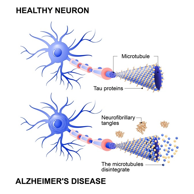
Alzheimer's disease is the change in tau protein that results in the breakdown of microtubules in brain cells. Mechanism of disease. two neurons: healthy cell and neuron with Alzheimers. Tau hypothesi. Image Credit: Designua / Shutterstock

For this effort the researchers looked at the brain tissues of a 74-year-old woman who had died. She was suffering from Alzheimer's disease. Alzheimer’s disease typically means progressive dementia or loss of memory and brain functions. It has been seen earlier that brains of individuals who study from this condition develop tangles of a protein called tau. These tangles or knots of protein spread throughout the brain. As they grow in numbers, they tend to worsen the symptoms. Tau tangles have been known to scientists and the medical fraternity for decades now. What was not known was how these tangles actually look like.

As a first, this team of researchers took detailed images of the tau inside the patient’s brain tissues. Using computer software they recreated how the tangles might look microscopically. While a picture of these proteins could not be something especially interesting or fascinating to most people, scientists believe that a detailed knowledge of how it looks to help develop drugs to control their growth. If the chemical structure of what is to be targeted is known clearly, it is but natural that drugs could be developed to fight it. According to Dr Sjors Scheres, one of the researchers, drugs aimed at the disease protein till date was akin to “shooting in the dark”. Now researchers have provided a concrete structure against which the weapons could be used more definitively. Why this study opens a “new era” in neurodegenerative disorders is clear. Other brain proteins that cause these diseases such as beta amyloid protein deposits in Alzheimer’s disease or alpha synuclein in Parkinson’s disease, can now all be clearly photographed and better understood explained researchers.

According to co researchers Dr Michel Goedert this was the first time a high resolution structure from human brain tissue was determined for these debilitating diseases. Not only drug development in these diseases, it could also help us understand the disease more clearly.

For this study the lab used a technique known as cryo-electron microscopy at 3.4–3.5 Å resolution. They mapped the finer details of the structure of the tau protein. In Alzheimer’s disease there are two types of abnormal proteins or neurofibrillary lesions as they are termed, in the brain. While tau filaments are formed within the nerve cells of the brain, beta amyloid proteins are formed outside the nerve cells in the form of filaments. Tau proteins in normal brains can help in nerve function. But among patients with Alzheimer’s they tend to get into clumps and tangles within the nerve cells. The images show that the filament cores of the tau protein are made up of two identical protofilaments that have residues 306–378 of tau protein. These form a cross-β/β-helix structure or coils around each other much like the DNA strands do.

Researcher Sjors Scheres said that this study can help show which parts of the filament formation of these tau proteins are actually important in the development of the disease and eventually could be drug targets.

Dr. Ananya Mandal

Written by

Dr. Ananya Mandal

Dr. Ananya Mandal is a doctor by profession, lecturer by vocation and a medical writer by passion. She specialized in Clinical Pharmacology after her bachelor's (MBBS). For her, health communication is not just writing complicated reviews for professionals but making medical knowledge understandable and available to the general public as well.

Citations

Please use one of the following formats to cite this article in your essay, paper or report:

  • APA

    Mandal, Ananya. (2023, July 10). Alzheimer’s disease specific brain deposits photographed for the first time in vivid detail. News-Medical. Retrieved on November 14, 2024 from https://www.news-medical.net/news/20170705/Alzheimers-disease-specific-brain-deposits-photographed-for-the-first-time-in-vivid-detail.aspx.

  • MLA

    Mandal, Ananya. "Alzheimer’s disease specific brain deposits photographed for the first time in vivid detail". News-Medical. 14 November 2024. <https://www.news-medical.net/news/20170705/Alzheimers-disease-specific-brain-deposits-photographed-for-the-first-time-in-vivid-detail.aspx>.

  • Chicago

    Mandal, Ananya. "Alzheimer’s disease specific brain deposits photographed for the first time in vivid detail". News-Medical. https://www.news-medical.net/news/20170705/Alzheimers-disease-specific-brain-deposits-photographed-for-the-first-time-in-vivid-detail.aspx. (accessed November 14, 2024).

  • Harvard

    Mandal, Ananya. 2023. Alzheimer’s disease specific brain deposits photographed for the first time in vivid detail. News-Medical, viewed 14 November 2024, https://www.news-medical.net/news/20170705/Alzheimers-disease-specific-brain-deposits-photographed-for-the-first-time-in-vivid-detail.aspx.

Comments

  1. jon robins jon robins United States says:

    This article is obviously not aware of the work done by the University of Calgary Faculty of medicine that has a video showing the neurofibrils tangles created by mercury that denudes the fiberals as the tubulin separates as a result of the electrical charge created by the mercury interaction causing the tangles.  We know also that the Ethyl mercury found in vaccines leaves the blood within 5 days of injection, but goes to the brain where it converts to INORGANIC mercury that is 50 times more toxic than methyl mercury and that vaccines contain polysorbate-80 that acts like a surfactant allowing heavy metals to easily cross the blood brain barrier including aluminum and mercury, especially the very toxic mercury from vaccines!  The idea of creating a drug to address this problem is ludicrous when we know what is creating the problem and addressing that ROOT CAUSE rather than obfuscating that TRUTH and looking at some toxic drug to address the problem that will most likely have side effects suppressing the immune system like all other drugs.  It's time to change the paradigm of drug company intervention and get REAL solutions to health problems of degenerative diseases and eliminate the man-made causations creating these issues.

The opinions expressed here are the views of the writer and do not necessarily reflect the views and opinions of News Medical.
Post a new comment
Post

While we only use edited and approved content for Azthena answers, it may on occasions provide incorrect responses. Please confirm any data provided with the related suppliers or authors. We do not provide medical advice, if you search for medical information you must always consult a medical professional before acting on any information provided.

Your questions, but not your email details will be shared with OpenAI and retained for 30 days in accordance with their privacy principles.

Please do not ask questions that use sensitive or confidential information.

Read the full Terms & Conditions.