Each complex structure of the human heart has specific roles that contribute to efficient cardiac function, and the interruption of any of these functions can lead to congenital disabilities such as congenital heart disease in children and cardiac diseases such as valvulopathies and cardiomyopathies in adults. However, despite the critical role of the heart in the human body, the organization and function of the cardiac structures and how they interact with one another remain poorly understood.
About the study
In the present study, the researchers used a single-cell RNA sequencing (scRNAseq) approach along with multiplexed error-robust fluorescence in situ hybridization (MER-FISH). This strategy allowed them to combine single-cell transcriptomes and spatial biology and visualize, analyze, and quantify the RNA transcripts of a large number of genes from a single cell.
They began by identifying the cell lineages that were part of the developing heart, which helped determine how the various cardiac cell types assemble into complex structures and coordinate to regulate the function of the human heart. The scRNAseq was conducted in replicates and analyzed for human hearts in various stages of growth, starting from nine weeks and going up to 16 weeks post-conception.
The obtained single cells, over 140 million in number, were transcriptionally categorized into five cell compartments: cardiomyocytes, endothelial, mesenchymal, neuronal, and blood. Within these cell compartments, analysis of gene markers helped identify 12 cell classes, with subsequent clustering analyses identifying 39 populations and 75 subpopulations of cells.
MER-FISH was then used to spatially map the heart cells and explore the cellular mechanisms through which the remodeling and morphogenesis of the heart, including the ventricular wall development, were directed. The organization of the cells identified using scRNAseq, especially during developmental periods such as the compaction of the myocardial wall, was explored using MER-FISH imaging.
The study then aimed to decipher the assembly of these specific cardiovascular cells into the cellular neighborhoods that come together to form the multi-cell structures that contribute to heart function. The scientists also explored the organizational and cellular complexity of specific regions, such as the ventricles, by exploring the cells within the ventricles that were identified, isolated, and mapped using MER-FISH. Additionally, mouse models were used to interrogate the interactions between cells through in vivo experiments, and pluripotent stem cells from humans were used to evaluate the same in in vitro experiments.
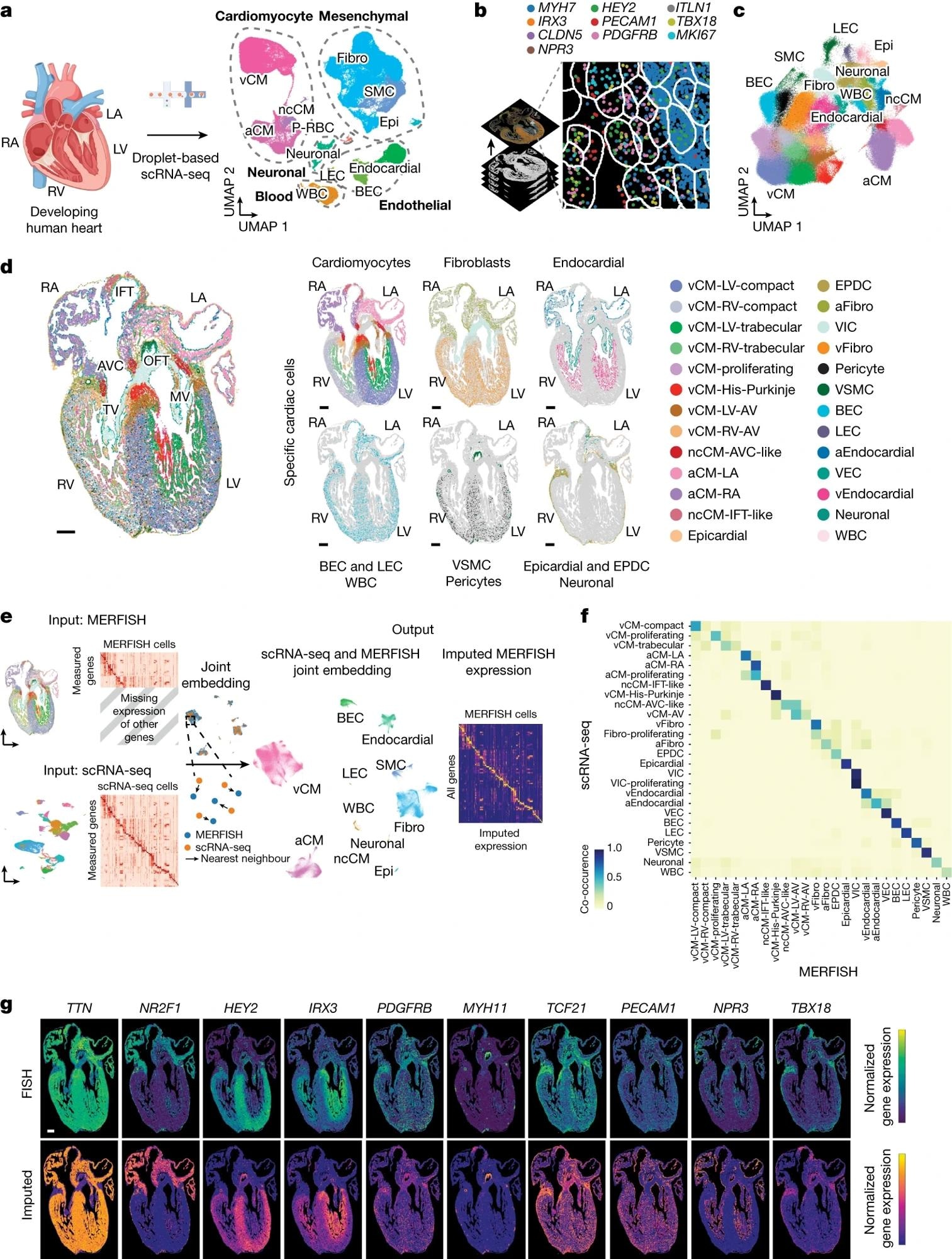 a, Left, schematic of experiment. Right, scRNA-seq identifies a diverse range of distinct cardiac cells that create the developing human heart as displayed by uniform manifold approximation and projection (UMAP) of ~143,000 cells. b, Schematic shows how 238 cardiac-cell-specific genes were spatially identified using MERFISH. Pseudo-coloured dots mark the location of individual molecules of ten specific RNA transcripts. c, Approximately 250,000 MERFISH-identified cardiac cells were clustered into specific cell populations as shown by UMAP and coloured accordingly in d. d, Identified MERFISH cells were spatially mapped across a frontal section of a 13 p.c.w. heart (left) and shown according to major cell classes (right). e, Joint embedding between MERFISH and age-matched scRNA-seq datasets enabled cell label transfer and MERFISH gene imputation. f, Co-occurrence heatmap shows the correspondence of cell annotations of MERFISH cells to those transferred from the 13 p.c.w. scRNA-seq dataset. g, Gene imputation performance was validated spatially by comparing normalized gene expression profiles of marker genes measured by MERFISH with the corresponding imputed gene expression profiles. Epi, epicardial; MV, mitral valve; P–RBC, platelet–red blood cell; TV, tricuspid valve. Scale bar, 250 µm (g). Illustration in a was created using BioRender (https://www.biorender.com).
Results
The findings revealed that various cardiac cell types belonged to specific subpopulations that were part of specific communities, with the functional specialization defined according to the anatomical region in which they were present and the cellular ecosystem. The cardiomyocyte lineages were the largest cell compartment identified using MER-FISH. The study also found that cells that belonged to non-cardiomyocyte cell compartments also underwent segregation into populations and subpopulations and contributed to the formation of specific structures and regions of the heart.
Cardiomyocyte subpopulations in the ventricular regions exhibited an ability to construct complex laminal structures in the ventricular wall and form cellular communities with other subpopulations of cardiac cells. Furthermore, the in vivo and in vitro experiments conducted to understand the interactions between cells revealed that the spatial organization of subpopulations of cardiac cells during the morphogenesis of the ventricular wall was conducted through various signaling pathways.
The study also found cardiac regions composed of spatially organized combinations of cell populations that segregated together, called cellular communities. These cellular communities varied in the number and types of cell populations, and within these communities, neighbors of each cardiac cell within a 150-micrometer radius were defined. These interacting populations of cells also had distinct cellular signaling pathways.
Conclusions
Overall, the study found that cardiomyocytes were the largest compartment of cell types in the developing heart, and all the cell types exhibited distinct structural and regional distributions in the heart. Specific cell populations also formed cellular communities in various combinations, with signaling pathways between the cell populations within the community defining their structure and function. The study helped in understanding the development of the complex structure of the human heart, providing potential avenues to treat structural heart diseases.
Journal reference:
- Farah, E.N., Hu, R.K., Kern, C., Zhang, Q., Lu, T., Ma, Q., Tran, S., Zhang, B., Carlin, D., Monell, A., Blair, A.P., Wang, Z., Eschbach, J., Li, B., Destici, E., Ren, B., Evans, S.M., Chen, S., Zhu, Q. and Chi, N.C. (2024). Spatially organized cellular communities form the developing human heart. Nature. DOI: 10.1038/s4158602407171z, https://www.nature.com/articles/s41586-024-07171-z
 AI-enabled stethoscope doubles detection of valvular heart disease