Light-sheet fluorescence microscopy is a state-of-the art imaging technique for live-cell imaging. Unlike traditional imaging techniques such as laser scanning and spinning disk confocal microscopy which are plagued by photobleaching and phototoxicity, light-sheet fluorescence microscopy can be used for imaging live specimens for days on end with minimal phototoxic effects.
This is because of the unique geometry utilized in light-sheet, in which the light path has been decoupled into separate excitation and detections pathways. In doing so, only the focal volume of interest is illuminated, reduces the amount of light that the sample is exposed to and thus reducing photobleaching and phototoxic effects.
In this webinar, Dr. Dane Maxfield discusses the benefits of light-sheet fluorescence microscopy as a gentle bio-imaging technique for imaging a wide range of biological samples with high spatial and temporal resolution. This webinar will focus around the Luxendo InVi-SPIM, an inverted light-sheet sold by Bruker Nano Surfaces. This webinar will focus around the Luxendo InVi-SPIM, an inverted light-sheet sold by Bruker Nano Surfaces.
To begin, we start by discussing the photon budget of the experiment. Since each fluorescent molecule has a limited lifetime and number of photons, it is important to maximize how these photons are used to get the most out of your imaging experiment. Traditional imaging techniques, like laser scanning confocal microscopy and spinning disk confocal microscopy, suffer from photobleaching and photoxicity because they excite all of the fluorophores in a sample in order to collect emission light from a small focal volume determined by the size of the pinhole.
In doing so, these photons are wasted by illuminating fluorophores outside the focal volume of interest. However in light-sheet, the unique geometry allows us to only illuminate the focal volume of interest. This in turn reduces the amount of excitation light the sample experiences, minimizing phototoxicity and photobleaching while maximising the acquisition speed.
To highlight this, Dr. Maxfield next presents some theoretical and experimental data showcasing the low photobleaching, low phototoxicity and high acquisition speed in light-sheet microscopy as compared to spinning disk and laser scanning confocal microscopy. This data, published by some of the pioneers of light-sheet microscopy, highlights the advantage of this technique for live-cell imaging.
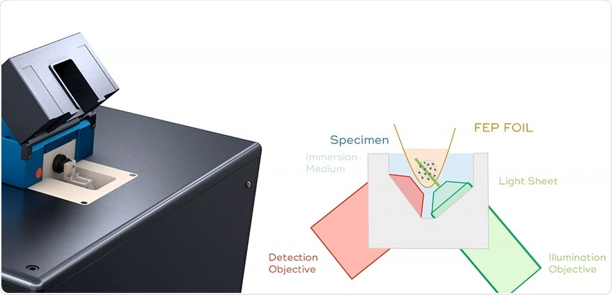
Dr. Maxfield wraps up the webinar by giving a detailed overview of the Luxendo InVi-SPIM, an inverted light-sheet microscope that has been specially designed for live imaging of small and extremely delicate biological samples. The InVi-SPIMs simple, easy sample mounting and full incubation chamber make this light-sheet system ideal for imaging organoids, spheroids, cell monolayers and mouse embryos at high spatial and temporal resolution. More details around the actual components, the internal workings and key specifications of the InVi-SPIM are detailed within the webinar.
To learn more about Bruker Nano Surfaces’ Luxendo product, the InVi-SPIM, and to see how using light-sheet microscopy could benefit your research, please click here to watch the webinar.