The LUMOS II ILIM is the fastest chemical imaging microscope, featuring a 2.2 x 2.0 mm2 field of view with 4.25 µm spatial resolution. Smart, application-specific workflows for tissue, particle, and tablet analysis simplify daily tasks.
Highlights
Meet the future of infrared imaging: LUMOS II ILIM
By integrating Infrared Laser Imaging (ILIM) into the LUMOS DNA, the system offers a groundbreaking shift from spectral analysis to image-based thinking. More than just a microscope, it functions as "Infrared Eyes," enabling sample characterization with unparalleled detail and IR imaging speed.
Meet the LUMOS II ILIM - the next generation of IR Laser Imaging
Video Credit: Bruker Optics
Start by discovering the dedicated ILIM applications ...
TissuePlusTM
Get essential information about the spatial biology of whole tissue microtome sections in a matter of minutes.
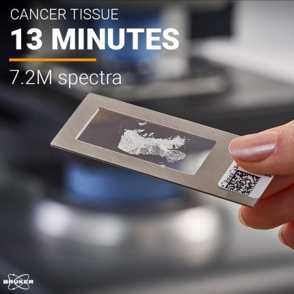
Image Credit: Bruker Optics
TabletPlusTM
In just a few minutes, analyze and deformulate pills and tablets. Get the chemical details of fillers, excipients, and APIs.
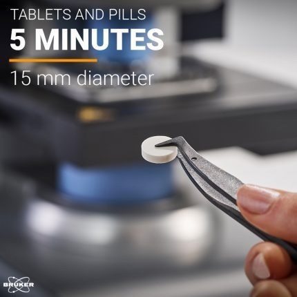
Image Credit: Bruker Optics
ParticlePlusTM
Get vital information about the chemical composition of particles and microplastics by analyzing entire filters in a matter of minutes.
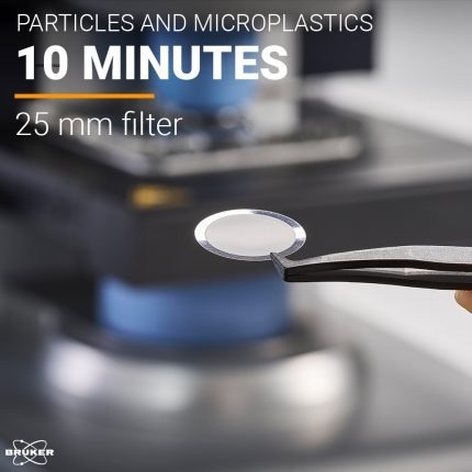
Image Credit: Bruker Optics
.. or develop your own IR imaging application
Technical features
- Patented spatial coherence reduction for high-quality IR images
- WinGuardTM technology ensures an open design while maintaining laser class 1 safety
- 4x lens objective (4.25 µm pixel size) with NA 0.6 and an extensive 2.2 mm x 2.0 mm field of view
- Exceptional IR imaging speeds, capturing up to 62,400 spectra per second across the full spectral range (249,600 spectra in 4 seconds @ 950-1800 cm-1)
- Fully automated hardware with intuitive push-button software design
- Validation plate for automated instrument qualification (OQ/PQ)
User benefits
- Up to 169x faster area scanning speed compared to FT-IR
- Routine-ready for application at scale
- No trade-off between measurement speed and spectral quality
- Truly developed for ease of use with dedicated workflows
We have been working in the field of tissue imaging and spectral pathology for over 20 years. One of the key barriers to clinical adoption is the long measurement times for full hyperspectral imaging of large areas of tissue. A sample set consisting of nearly 1500 prostate tissue cores, that took 3 months to measure on our old instrument, was measured in just two days on the Bruker ILIM system. This is a game changer!
Peter Gardner, Ph.D., Professor of Analytical and Biomedical Spectroscopy, University of Manchester