This article is based on a poster originally authored by Chitra Seewooruthun and contains work authored by Austin, M. Burschowsky, et al., and Rebecca Newton, et al. This poster was originally presented at ELRIG Drug Discovery 2023 in affiliation with the Institute of Precision Health and the University of Leicester.
This poster is being hosted on this website in its raw form, without modifications. It has not undergone peer review but has been reviewed to meet AZoNetwork's editorial quality standards. The information contained is for informational purposes only and should not be considered validated by independent peer assessment.

A binuclear manganese metalloenzyme known as arginase 2 (ARG2) catalyzes the conversion of L-arginine to L-ornithine and urea. Acute myeloid leukemia (AML) and other tumor microenvironments with dysregulated expression of ARG2 produce an immunosuppressive niche that essentially makes the tumor “invisible” to the host immune system.

Local L-arginine levels are concurrently depleted by increased ARG2 expression and release, which suppresses T-cell-mediated anti-tumor immunological responses, including reduced T-cell proliferation and T-cell receptor down-regulation.1
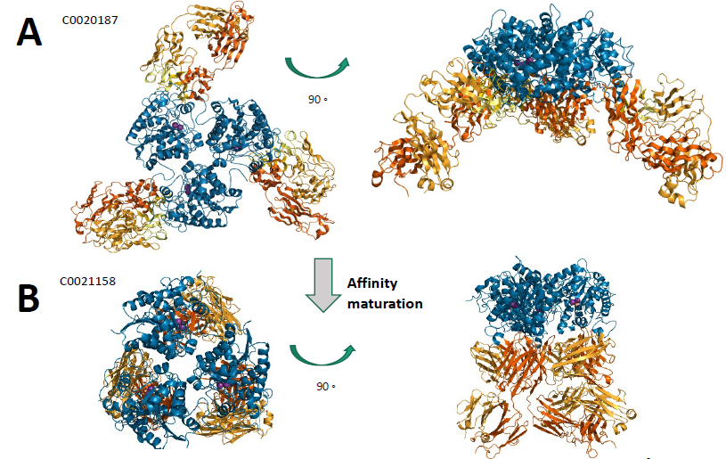
The parent inhibitory antibody C0020187 (Figure 1A) was recombined to create unbiased libraries using antibody chain shuffling and a staggered-extension procedure (StEP). Pool maturation was then performed to simultaneously optimize seven leads of interest.2
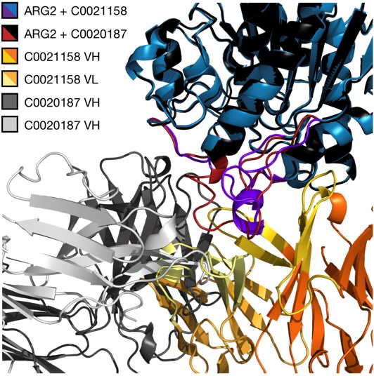
The significant structural alterations from parent to affinity-matured antibodies bound to ARG2, with a significant reorientation of the binding surface caused by the extensive sequence modifications, translated into significant improvements in binding characteristics and inhibitory efficacy (Figure 1 and 2).

Figure 1. Co-crystal structure of the Fab in complex with trimeric ARG2. A) C0020187 (PDB 6SS5 – 1.7 Å) (B) C0021158 (PDB 6SS2 –2.4 Å).

Figure 2. Epitope shift driven by changes in CDRH1.
Complete inhibition of ARG2 is achieved by the affinity-matured antibody C0021158 (Figure 1B), which also successfully restores T-cell proliferation in vitro. The lack of overlap between the epitope and the substrate binding cleft suggests an allosteric mechanism. When C0021158 binds, ARG2 experiences significant confirmational changes.
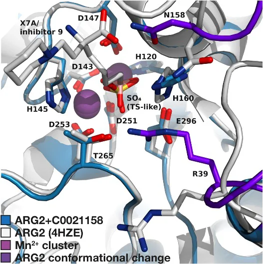
The C0021158 epitope results in more minute modifications to the active site of the enzyme, where an inward reorientation of Arg39 sterically prevents L-arginine from binding and modifies the pKA of a catalytic residue (His160) (Figure 3). In silico molecular docking simulations and isothermal calorimetry tests using an L-arginine mimetic3 provide support for this process.

Figure 3. Fab C0021158 binding rotates Arg39 into the active site of ARG2.
References
- Martí i Líndez et al. Mitochondrial arginase-2 is a cell-autonomous regulator of CD8+ T cell function and antitumor efficacy. JCI Insight. 2019;4:e132975.
- Chan et al., Extensive sequence and structural evolution of Arginase 2 inhibitory antibodies enabled by an unbiased approach to affinity maturation. Proc Natl Acad Sci US A. 2020 Jul 21;117(29):16949-16960.
- Austin et al. Structural and functional characterization of C0021158, a high-affinity monoclonal antibody that inhibits Arginase 2 function via a novel non-competitive mechanism of action. MAbs. 2020 Jan-Dec;12(1):1801230.
Structure-based drug design - RET-driven lung adenocarcinoma
Rearranged during transfection (RET) protein kinase is a type of receptor tyrosine kinase that plays a crucial role in cell proliferation, survival, and differentiation.
It has recently been determined that activating gene fusions of the receptor tyrosine kinase, or RET, are responsible for 1-2% of all non-small cell lung malignancies (NSCLC)1-3. Vandetanib and cabozantinib are clinically authorized for this indication; however, RET inhibition is a secondary pharmacology.4-5
The primary pharmacology of these agents, which inhibit KDR (or VEGFR2), results in dose-limited adverse effects, limiting their effectiveness in treating NSCLC.
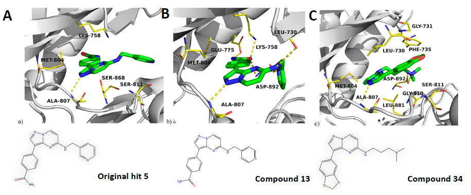
More selective RET inhibitors that show no KDR inhibitory effect in vivo have drawn much attention in light of the reported clinical toxicity. Drugs like LOXO-292 and BLU-667 are now undergoing clinical trial evaluation. Several inhibitors of RETV804M kinase, the predicted drug-resistant mutant of RET kinase, have been developed due to concentrated library and virtual screening, hit expansion, and rational design (Figure 1).

Figure 1. A) Docked model of early hit 5 bound to RETV804M. (B) Crystal structure of 13 bound to RETV804M (PDB 6I83). (C) Crystal structure of optimized lead 34 bound to RETV804M (PDB 6I82). In all panels, RETV804M is shown as grey cartoons, key residues as yellow sticks, and the respective ligand as green sticks.
These agents provide a possible supplement to RET inhibitors that are now undergoing clinical investigation since they do not inhibit the wild type (wt) isoforms of RET or KDR (Table 1).
Table 1. Optimization of hit 5.
| |
Original hit 5 |
Compound 13 |
Compound 34 |
| RETV804M ENZYME IC50 |
20 nM |
51 nM |
19 nM |
| RET Selectivity (enzyme) |
3.7 X |
13 X |
16 x |
| KDR selectivity (enzyme) |
110 x |
19 x |
>410 x |
| RETV804M cell IC50 |
4.4 μM |
3.9 μM |
120 nM |
| RET selectivity (cell) |
0.8 x |
0.8 x |
32 x |
| KDR selectivity (cell) |
2.3 x |
1.9 x |
76 x |
References
- Martí I Líndez et al. Mitochondrial arginase-2 is a cell-autonomous regulator of CD8+ T cell function and antitumor efficacy. JCI Insight. 2019;4:e132975
- Chan et al., Extensive sequence and structural evolution of Arginase 2 inhibitory antibodies enabled by an unbiased approach to affinity maturation. Proc Natl Acad Sci U S A. 2020 Jul 21;117(29):16949-16960
- Austin et al. Structural and functional characterization of C0021158, a high-affinity monoclonal antibody that inhibits Arginase 2 function via a novel non-competitive mechanism of action. MAbs. 2020 Jan-Dec;12(1):1801230
- Kohno et al . KIF5B-RET fusions in lung adenocarcinoma. Nat. Med. 2012, 18 (3), 375– 377
- Takeuchiet al. RET, ROS1 and ALK fusions in lung cancer. Nat. Med. 2012, 18 (3), 378– 381
- Lipson et al. Identification of new ALK and RET gene fusions from colorectal and lung cancer biopsies. Nat.Med. 2012, 18 (3), 382– 384
- Drilon et al. Cabozantinib in patients with advanced RET-rearranged non-small-cell lung cancer: an open-label, single-centre, phase 2, single-arm trial. Lancet Oncol. 2016, 17 (12), 1653– 1660
- Yoh et al. Vandetanib in patients with previously treated RET-rearranged advanced non-small-cell lung cancer (LURET): an open-label, multicentre phase 2 trial. Lancet Respir. Med. 2017, 5 (1), 42– 50
- Newton et al., Extensive sequence and structural evolution of Arginase 2 inhibitory antibodies enabled by an unbiased approach to affinity maturation, ACS Medicinal Chemistry Letters 2020 11 (4), 497-505
Last Updated: Nov 18, 2024