Angiosarcoma (AS), which is a soft tissue tumor that affects the inner layer of blood and lymph vessels, is often asymptomatic in the early stages. Most of the sub-types of this disease manifest only when it has reached a serious level or has spread to other parts of the body.
Some types, such as cutaneous angiosarcoma, present with symptoms such as a rash and staining on skin surfaces, which may be misinterpreted as normal skin ailments. These characteristics of the disease can lead to progression of the malignancy, and early diagnosis and treatment is key to the better patient outcomes.

Angioma or hemangioma a vascular tumor composed of small blood vessels lined by endothelial cells. Cavernous angioma. Image Credit: Shutterstock
Methods of Diagnosis
The diagnostic techniques used to confirm a diagnosis of angiosarcomas are very similar to those used for other cancers. After a primary physical examination, if the physician suspects the presence of a tumor, he might suggest diagnostic imaging techniques, through which he can clarify the condition. Magnetic resonance imaging (MRI), computed tomography (CT) and positron emission tomography (PET) are the common imaging tests used in the diagnosis of a tumor. Biopsy is an important element in the diagnosis of cancer to confirm the malignant nature of the cells following imaging tests.
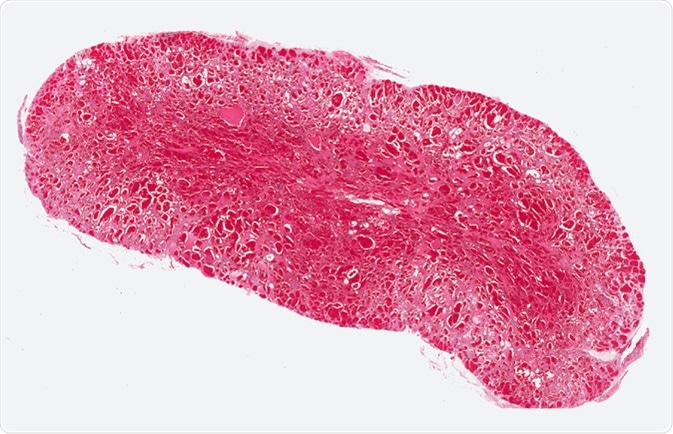
Angiosarcoma
Physical Examination
This is the first step in diagnosing any illness. In the case of AS, the findings by physical examination might not be remarkable. However, several few clues may be evident, which prompt the doctor to suggest further screenings.
For breast AS, the doctor or specialist nurse looks for a bulge or enlargement in the breast area, under arms, and the base of the neck. Similarly, each subtype may have a few manifestations depending on the affected area.
Image Testing
- MRI scan: Magnetic resonance imaging or MRI scan is an imaging technique that offers a definite screening of the tissues. In addition to imaging non-specific mass of tumors, it helps in the planning of surgery by evaluating the response of the tumors to radiotherapy and chemotherapy. Vascular lesions can be diagnosed by this technique because of its capability to detect vascular channels and spaces.
- PET scan: Positron emission tomography or PET scan is used to determine the metabolic activity of tumors. Combined with CT, this technique helps detect cancer as well as classify it by stage or grade. The technique is also used to confirm the location of the tumor for biopsy and to evaluate the efficiency of treatments.
- CT scan: CT or computed tomography provides an image of the tumor to detect the shape and size of the growth. It is also used to guide a needle while removing tissues for biopsy (CT-guided biopsy).
- Mammogram: This technique is used to detect ASs of the breast. A mammogram is typically an X-ray technique used to scan the breasts of women, who are suspected to have cancer. It reveals details such as thickening of the skin or presence of a superficial mass in the breast. But few studies suggest that nearly 33% of the patients who were suspected to have an AS of the breast were not identified by this method.
- Ultrasound scanning: Sonography or an ultrasound is another imaging technique, which creates images of organs inside the body with the use of high-frequency sound waves. If a person subjected to a diagnosis of cancer is under the age of 35 years, an ultrasound is usually used in preference to a mammogram.
The exact location of the tumor in the body can be detected through this technique. The sound waves used pass through the body and bounce back when they hit an organ. These waves are then received by a device called transducer and transformed into an image.
Histopathologic Verification
Verification of a sample of cells affected by tumor under a microscope can help find the grade of the condition. Normally, lesions that look less malignant are classified as low grade and might be well differentiated, whereas poorly differentiated and malignant lesions are classified as high grade.
AS has three grades: I (well differentiated), II (moderately differentiated), and III (poorly differentiated).
Biopsy: A proper and well-defined diagnosis of any kind of AS is made using a biopsy. The term ‘biopsy’ can be defined as a tiny sample of the cell or tissue that has been taken from the tumor. A pathologist verifies this biopsy under a microscope to find the stage of growth of the tumor.
To obtain proper sections of tissues that are helpful in determining the grade and histologic type, larger samples of tissues are needed. Fine-needle aspiration (FNA) and core-needle biopsies (CNB) are two tools used in these types of tests.
Determining the stage or grade of the disease is very important for devising proper treatment strategies. In most cases, angiosarcoma is an aggressive and fast spreading tumor and it rarely has a contradictory character with slower spreading and being less malignant. The decreased life expectancy that the disease causes with an average of 5 years is the factor that makes “early and efficient diagnosis” vital.
Further Reading
Last Updated: Feb 26, 2019