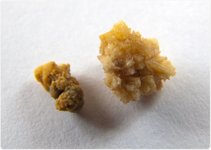
Ureterolithiasis or ureteric calculi are stones that form in or travel down to the ureters, which are the slender muscular tubes that connect the kidneys to the urinary bladder. These tubes are physiologically constricted at 3 points along their lengths, namely at the ureteropelvic junction, entry into the pelvis as they cross the bifurcation of the common iliac artery and the ureterovesical junction.
As a consequence, these points are the commonest sites where ureteric calculi may become impacted. This may cause several signs and symptoms that may be site-specific as well as related to the degree of urinary obstruction.

Credit: remik44992/Shutterstock.com
Patients may present with nausea, vomiting, blood in the urine and pain in the back, flank and lower abdomen. This pain is usually colicky in nature, which means that it presents in a pattern of waves owing to the peristalsis of the ureters. However, it may also be constant.
Typically, the pain is abrupt in onset and may be confused with other abdominal pathologies depending on exactly where within the abdomen it radiates to. Right-sided pain may be confused, for example, with appendicitis (i.e. inflammation of the appendix). There are several different types of calculi and the etiology surrounding the development of ureteric calculi may be multifactorial.
Types of ureteric stones
Based on chemical composition, there are four main types of stones that may be present in the ureters. These are calcium, struvite, uric acid and cysteine calculi. Nearly 8 in every 10 ureteric stones will be found to be composed predominantly of calcium. Struvite stones are those that are composed of magnesium ammonium phosphate and these account for around 15% of cases, while uric acid and cysteine stones account for approximately 6% and 2%, respectively. The etiological factors for these different types of stones vary.
Etiology
Stone formation is believed to be driven by a series of events that follow, at least in part, inadequate fluid intake, which results in a directly proportional lowered urinary output. This, in turn, gives rise to a high solute concentration in the urine that provides favorable condition for the development of urinary stones. This cascade may be regarded as the most important environmental cause of stone formation. Notwithstanding this observation, there is also a link to metabolic dysfunction and the development of urinary stones. In particular, hypercalciuria (i.e. increased urinary calcium) has been implicated.
Hypercalciuria may arise due to many different reasons. It has been shown to be associated with increased absorption of calcium within the intestines. This increased absorption may be a consequence of failure in the mechanisms that regulate the absorption of calcium or there may be a dietary excess of calcium. Despite the latter, evidence does not suggest restricting calcium intake in persons prone to developing calcium stones.
Instead a diet low in protein and salt is recommended as opposed to one low in calcium. Another cause of hypercalciuria may berenal tubular dysfunction as it pertains to the ability of the kidneys to filter and reabsorb calcium.
Chronic urinary tract infections (UTIs), which may be associated with anatomical irregularities within the urinary system, have been shown to be the culprits inthe development of struvite stones. The organisms responsible for these infections and struvite stone formation are those such as Pseudomonas, Proteus and Klebsiella species, which convert urea into ammonia. This product subsequently combines with magnesium and phosphate to crystalize and form a stone.
Acidic urine, or a diet rich in purines, and/ or malignancies have all been implicated in the formation of uric acid stones. Cysteine stones on the other hand are associated with metabolic defects causing high levels ofamino acids such as cysteine and ornithine, and the inability of the renal tubules to reabsorb them. Aside from diet, infections and derangements in anatomy, physiology or metabolism, stones may also form directly from some medications or indirectly as a result of high concentrations of their breakdown products.
References
- https://uroweb.org/
- http://pmj.bmj.com/content/postgradmedj/27/305/128.full.pdf
- https://www.ncbi.nlm.nih.gov/pmc/articles/PMC2600100/
- https://radiopaedia.org/articles/ureter
- https://www.ncbi.nlm.nih.gov/pmc/articles/PMC2518455/
Further Reading
Last Updated: Dec 28, 2022