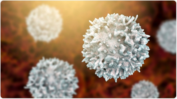
The pathophysiology of severe acute respiratory syndrome coronavirus 2 (SARS-CoV-2) involves a highly aggressive inflammatory response that primarily damages the respiratory tract. Aside from the viral infection itself, the host response has also been found to play a key role in determining the severity of coronavirus disease 2019 (COVID-19).

Image Credit: Kateryna Kon/Shutterstock.com
How does SARS-CoV-2 infect cells?
Studies on the pathogenesis of the novel coronavirus have confirmed that SARS-CoV-2 uses angiotensin-converting enzyme 2 (ACE2) receptors to enter cells. Although ACE2 is expressed in most tissues throughout the body, its highest expression can be found in the kidney, endothelium, heart, and lungs.
In synergy with ACE2, SARS-CoV-2 must also interact with transmembrane protease 2 (TMPRSS2) to enter the cell. Upon entry into the cells, SARS-CoV-2 undergoes active replication until the host cell undergoes pyroptosis, which is a highly inflammatory type of programmed cell death.
A dysfunctional immune response to SARS-CoV-2
Pyroptotic cells will release several different damage-associated molecules including adenosine triphosphate (ATP), nucleic acids, and apoptosis-associated speck-like (ASC) oligomers. Neighboring endothelial and epithelial cells, as well as alveolar macrophages within the lungs, will recognize the presence of these molecules, thereby triggering an inflammatory response that involves pro-inflammatory cytokines and chemokines like interleukin (IL)-6, IL-10, macrophage inflammatory protein 1a (MIP1a) and MIP1b.
A positive feedback loop between these released proteins and certain immune cells like monocytes, macrophages, and T cells will be created to promote the inflammatory response.
When the immune system is in disarray, this pro-inflammatory feedback loop will cause an excessive infiltration of inflammatory cells to the site of injury. Since SARS-CoV-2 primarily uses ACE2 to enter lung cells, there is a reduced function of ACE2 to perform its normal functions within the renin-angiotensin system (RAS). This dysfunction of the RAS disrupts the normal balance that exists between the blood pressure and electrolyte levels and enhances the vascular permeability of the airways, thereby further enhancing the release of inflammatory cells to infected lung cells.
Taken together, pulmonary edema and pneumonia arise, which not only causes damage to the lung infrastructure but can also lead to widespread inflammation and damage in other organs.
T cell immunity
Once an individual becomes infected with SARS-CoV-2, the virus will remain in its incubation period for between 4 and 5 days before the patient begins to develop symptoms. Approximately one week after COVID-19 symptoms begin, both B and T cell responses can be detected in the blood.
Early studies conducted on some of the first COVID-19 patients found that mononuclear cells, which most likely included monocytes and T cells, accumulated within the lungs, whereas low levels of hyperactive T cells were identified in the peripheral blood. The presence of T cells in such low levels within the blood suggests that rather than remain within the bloodstream, T cells travel from the blood into the infected organs to mitigate the immune response.
Since SARS-CoV-2 shares a 79% resemblance to the genetic profile of SARS-CoV that infected patients between 2003 and 2004, researchers believe that infection by SARS-CoV-2 also initiates a TH1 cell response.
More specifically, this response involves the there is a massive T cell response to the acute infection, in which CD4+ and CD8+ T cells are predominantly involved. While this early T cell response appears to be protective, the ability of this specific immune response to prevent infection in humans has not been fully evaluated.

Image Credit: Kateryna Kon/Shutterstock.com
B cell response
Humoral immunity depends on the ability of B cells to successfully transform into plasmocytes and produce antibodies. The production of antibodies to COVID-19 is largely dependent upon the response by T follicular helper cells, which typically occurs one week after COVID-19 symptoms begin.
To date, a total of 19 neutralizing antibodies are produced by B cells in COVID-19 patients. Whereas nine of the identified neutralizing antibodies to SARs-CoV-2 bind to the receptor-binding domain (RBD) of the spike (S) protein of this virus molecule, eight other neutralizing antibodies target the N-terminal region of the upper S protein and the remaining two antibodies bind to other nearby regions.
The COVID-19 antibody response has been identified to begin between 4 and 8 days after symptoms begin; however, the neutralizing activities of these antibodies do not begin until at least 2 weeks after symptom onset.
These antibodies have the potential, with the assistance of memory B cells, to prevent reinfection if a patient encounters SARS-CoV-2 again in the future.
Current evidence, suggests that neutralizing antibody activity declines post-infection and offers short-term humoral immunity through stable neutralizing antibodies for at least 5 to 7 months (although this number has varied with different studies and individuals).
Several monoclonal antibodies have been developed from the memory B cells of former COVID-19 patients as potential therapies. Some of these treatments have been approved for emergency use by the FDA.
Although the long-term stability of these neutralizing antibodies has not yet been confirmed, they have the potential, with the assistance of memory B cells, to prevent reinfection if a patient encounters SARS-CoV-2 again in the future. Several monoclonal antibodies
have been developed from the memory B cells of former COVID-19 patients as potential therapies.
Although the neutralization capabilities of certain antibodies are believed to be effective against SARS-CoV-2 and thereby have protective effects, B cells can also produce non-neutralizing antibodies.
The release of non-neutralizing antibodies could potentially initiate a process known as antibody-dependent enhancement (ADE) of the disease. Rather than offer protective effects against SARS-CoV-2, these alternative non-neutralizing antibodies could instead enhance SARS-CoV-2 infection by ADE.
This was a concern when developing SARS-CoV-2 vaccines. However, evidence from trials of the vaccines in use suggests no sign of ADE responses.
Conclusion
The immune response to SARS-CoV-2 is crucial to not only understanding the pathogenesis of this highly contagious virus, but also the design and evaluation of vaccines. As SARS-CoV-2 continues to travel around the world, the virus will continue to undergo different mutations is inevitable.
Mutations that result in altered S protein configurations could become resistant to some monoclonal antibodies, thereby rendering them ineffective. Another important aspect of immunity estimations is their role in determining which future pandemic control measures, such as social distancing and mask requirements, are employed.
References
- Tay, M. Z., Poh, C. M., Renia, L., et al. (2020). The trinity of COVID-19: immunity, inflammation, and intervention. Nature Reviews Immunology 20; 363-374. doi:10.1038/s41577-020-0311-8.
- “How does a SARS-CoV-2 Virion Bind to ACE2?”
- Grifoni, A., Weiskopf, D., Ramirez, S. I., et al. (2020). Targets of T Cell Responses to SARS-CoV-2 Coronavirus in Humans with COVID-19 Disease and Unexposed Individuals. Cell 181(7); 1489-1501. doi:10.1016/j.cell.2020.05.015.
- Arvin, A. M., Fink, K., Schmid, M. A., et al. (2020). A perspective on potential antibody-dependent enhancement of SARS-CoV-2. Nature 584; 353-363. doi:10.1038/s41586-020-2538-8.
- “Potent neutralizing antibodies target new regions of coronavirus spike” – National Institutes of Health
Further Reading
Last Updated: Mar 30, 2021