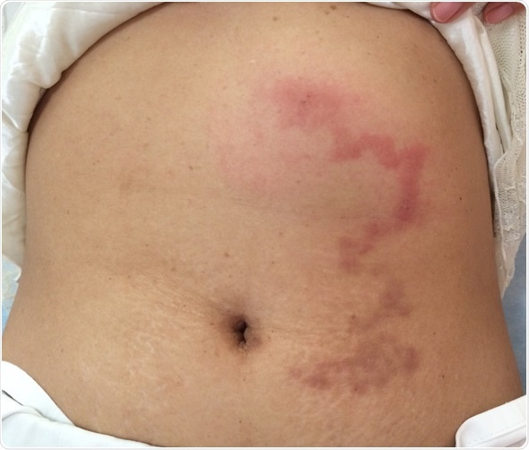
Cutaneous larva migrans is a creeping skin eruption with a serpentine single-track rash. Most common in those who live in or visit the tropical and subtropical areas of the world, it is caused by a helminthic infestation.
This parasitic skin disease is due to direct contact with the larva of the hookworm (Ancylostoma species), which has its definitive host in cats or dogs.

Cutaneous larva migrans at abdominal wall. Image Credit: TisforThan / Shutterstock
In a few cases it is caused by the dog tapeworm (Strongyloides) larva. It is usually acquired by bare skin contact with the soil in places where the soil is contaminated with the feces of these animals.
About 700 million people are estimated to suffer from cutaneous larva migrans each year. Most of these are children, sunbathers on tropical beaches, or outdoor workers. Geographically, its incidence is highest in the south of the USA, southern and eastern Africa and South-East Asia.
How it is Acquired
The parasite has its definitive life cycle in the dog or cat. When these animals defecate, the eggs of the hookworm are passed on to the soil. Here they hatch and develop into the infective stage. These larvae are capable of penetrating bare skin, because of their protease enzymes. These enzymes help them enter through cracks or hair follicles, or even through intact skin. They may leave a reddish, itchy papule or vesicle at the site of entry, which is usually between the toes.
The larvae then travel through the skin, leaving the characteristic tortuous lines that appear within 2 weeks of infestation. They may lengthen as much as 2 cm a day. Finally, the larvae die in the epidermis, unable to complete their life cycle. They produce persistent itching, and sometimes pain and swelling around the rash. Fever may also be present in some cases.
Complications
The skin may be so itchy that it is frequently scratched, leading to secondary infection. Pneumonitis may rarely develop due to larval migration through the lungs. This is referred to as Loeffler’s syndrome and is associated with massive larval infestation. Eosinophilic enteritis is another rare complication.
Diagnosis and Management
The characteristic appearance of the rash coupled with the history of exposure of bare skin to an infested soil or sandy surface is usually diagnostic. Sometimes the rash may look different due to the formation of vesicles, or following fungal or secondary bacterial infection. Microscopy reveals the typical filariform hookworm larvae.
Blood tests show eosinophilia and high serum Immunoglobulin E (IgE) levels.
The larvae usually die in a few weeks or months, even without treatment. However, ivermectin, thiobendazole or albendazole are commonly used medications to eradicate the parasitic larvae.
Prevention
Prevention is better than cure. The best way to prevent cutaneous larva migrans is to avoid direct proximity between the bare skin and the ground in places where infection risk is likely.
Thus a beach towel should be used before sitting or lying on the ground. Footwear should be worn on the beach in suspected places and beaches should be kept free of animal feces.
References
Further Reading
Last Updated: Feb 26, 2019