From RevvityReviewed by Olivia Frost
This article is based on a poster originally authored by Yongyang Huang, Ning Guo, Mackenzie Pierce, Leo Chan, Bo Lin and Yi Yang, which was presented at ELRIG Drug Discovery 2024 in affiliation with Revvity Health Sciences, Inc., and Novartis.
This poster is being hosted on this website in its raw form, without modifications. It has not undergone peer review but has been reviewed to meet AZoNetwork's editorial quality standards. The information contained is for informational purposes only and should not be considered validated by independent peer assessment.

Abstract
Chimeric antigen receptor (CAR) T cell therapy is a novel cellular therapeutic approach for cancer patients, including those with B-cell malignancies. A critical step in the CAR-T cell production process is effectively delivering CAR genes into primary T cells, which can be achieved through viral vectors or non-viral methods.
It is imperative to perform analytical tests to detect and monitor cell proliferation, cell health status, and CAR gene expression to evaluate and select effective CAR gene delivery methods and processes. In this work, we developed an image-based method using the Cellaca® PLX Image Cytometer to quickly count T-cells, measure viability, assess apoptotic cell health, and identify CAR expression.
Using this new methodology, we compared different CAR gene delivery methods, primarily focusing on non-viral methods involving electroporation. Cell viabilities were monitored daily using acridine orange / propidium iodide (AO/PI) stain and its respective dual fluorescent assay. Preliminary results showed that viabilities for all SupT1 samples decreased significantly to ~50% by day 1 following electroporation, in comparison to un-transduced SupT1 control samples, which maintained ~90%+ viabilities.
These results confirmed that the introduction of plasmids, rather than the electroporation process itself, induced apoptosis and, eventually, cell death. Annexin V / PI and Caspase-3 / RubyDead cell health assays were also tested, and results indicated that most cell death following electroporation was likely the result of apoptotic cells transitioning to the point of no return–cell death. Transduced SupT1 samples fully recovered, as their viabilities increased to ~90%+ by day 5 of the study. Lastly, samples were stained with APC-conjugated specific anti-CAR antibody, and SupT1 CAR expression levels were measured using the Cellaca® PLX, and results were confirmed using a flow cytometer.
Utilizing this image-based method, we could monitor CAR expression in SupT1 cell samples on day 2, 5, 7 and compare CAR expression levels among different gene delivery methods (viral vectors or non-viral methods). With the advantages of ease of use, visual verification with captured cell images, and higher-throughput capability, the Cellaca® PLX Image Cytometer may be potentially used as a convenient benchtop system for rapid assessment of the quantity and quality of CAR-T cells, which may ultimately improve the productivity of development and manufacturability of CAR-T products.
General process of CAR-T cell production
Chimeric antigen receptor (CAR) T cell therapy involves engineering T cells to attack tumor cells by specifically binding to tumor antigens.
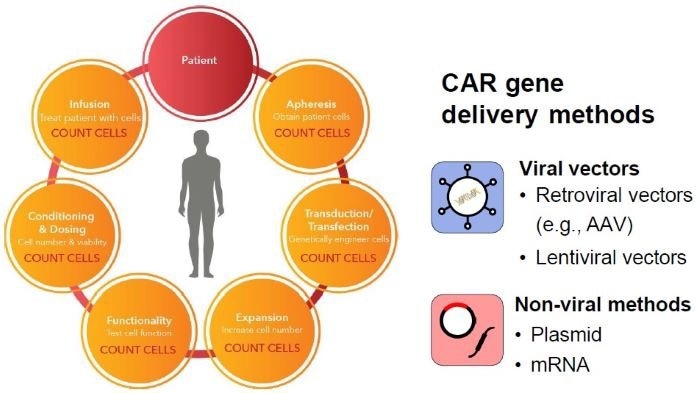
Image Credit: Image courtesy of Yongyang Huang et al., in partnership with ELRIG (UK) Ltd.
Workflow for CAR gene delivery, cell expansion and characterization

Image Credit: Image courtesy of Yongyang Huang et al., in partnership with ELRIG (UK) Ltd.
CAR-T cell viability results with premixed duo fluorescent AO/PI dyes
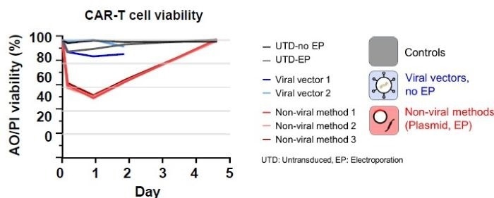
Image Credit: Image courtesy of Yongyang Huang et al., in partnership with ELRIG (UK) Ltd.
- Electroporation itself does not cause a significant drop in cell viability
- Viral vectors have a minor impact on cell viability
- Cell viability using non-viral methods drops to ~60% at ~4 hours and ~50% by day 1
Monitoring apoptosis using Annexin V / PI and Caspase-3 / RubyDead assays
Annexin V: Detects early-stage apoptosis
Caspase-3: Detects late-stage apoptosis
PI or RubyDead: Detects dead cells
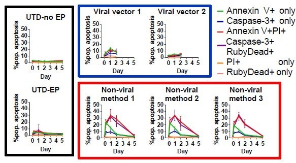
Image Credit: Image courtesy of Yongyang Huang et al., in partnership with ELRIG (UK) Ltd.
- Electroporation (EP) causes a small increase in apoptosis (~10%).
- Introducing a plasmid or a viral vector may induce significant apoptosis.
Transition from early- to late-stage apoptosis, and eventually cell death
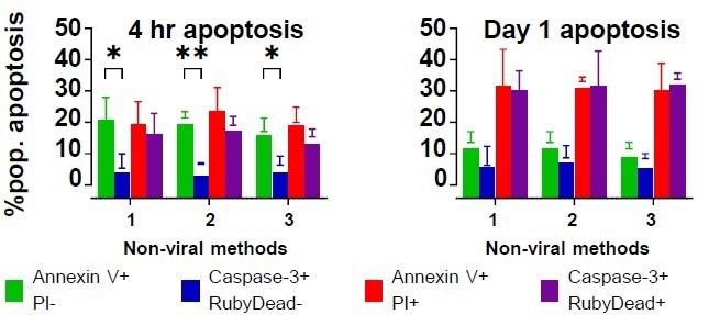
Image Credit: Image courtesy of Yongyang Huang et al., in partnership with ELRIG (UK) Ltd.
- At ~4 hours, the difference in the percentage of early- and late-stage apoptotic cells is ~15%.
- On day 1, the early-stage apoptotic cell population drops, while the late-stage apoptotic and dead cell populations increase
Identification of CAR expression
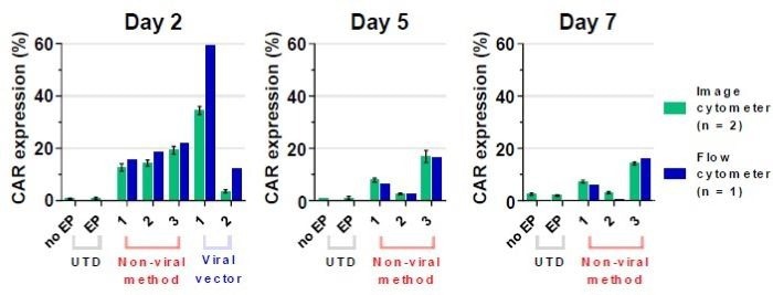
Image Credit: Image courtesy of Yongyang Huang et al., in partnership with ELRIG (UK) Ltd.
- CAR expression results between image and flow cytometers are comparable.
- Except for viral vectors 1 and 2 on day 2.
8. Multiplexed apoptosis assay

Image Credit: Image courtesy of Yongyang Huang et al., in partnership with ELRIG (UK) Ltd.
- Multiplexed apoptosis assay can provide an additional examination on whether the Annexin V+ cells are also Caspase-3+.
Multiplexed CAR expression and apoptosis

Image Credit: Image courtesy of Yongyang Huang et al., in partnership with ELRIG (UK) Ltd.
- Multiplexed CAR expression and apoptosis assay can detect the percentage of CAR+ cells that are or are not late-stage apoptotic.
Summary
- Non-viral methods for CAR gene delivery resulted in a significant decrease in cell viability following electroporation.
- Most cells entered the apoptotic pathway following electroporation, eventually entering the point of no return – cell death.
- CAR expression can be characterized using image cytometry, with results confirmed through flow cytometry.
- Image cytometry-based multiplex assays can provide rapid measurements for the following: direct cell counting, viability, apoptosis, and CAR expression
About Revvity
Revvity provides health science solutions, technologies, expertise, and services that deliver complete workflows from discovery to development, and diagnosis to cure.
Revvity is pushing the limits of what’s possible in healthcare, with specialized focus areas in translational multi-omics technologies, biomarker identification, imaging, prediction, screening, detection and diagnosis, informatics and more.
About ELRIG (UK) Ltd.
The European Laboratory Research & Innovation Group (ELRIG) is a leading European not-for-profit organization that exists to provide outstanding scientific content to the life science community. The foundation of the organization is based on the use and application of automation, robotics and instrumentation in life science laboratories, but over time, we have evolved to respond to the needs of biopharma by developing scientific programmes that focus on cutting-edge research areas that have the potential to revolutionize drug discovery.
Comprised of a global community of over 12,000 life science professionals, participating in our events, whether it be at one of our scientific conferences or one of our networking meetings, will enable any of our community to exchange information, within disciplines and across academic and biopharmaceutical organizations, on an open access basis, as all our events are free-of-charge to attend!
Our values
Our values are to always ensure the highest quality of content and that content will be made readily accessible to all, and that we will always be an inclusive organization, serving a diverse scientific network. In addition, ELRIG will always be a volunteer led organization, run by and for the life sciences community, on a not-for-profit basis.
Our purpose
ELRIG is a company whose purpose is to bring the life science and drug discovery communities together to learn, share, connect, innovate and collaborate, on an open access basis. We achieve this through the provision of world class conferences, networking events, webinars and digital content.
Sponsored Content Policy: News-Medical.net publishes articles and related content that may be derived from sources where we have existing commercial relationships, provided such content adds value to the core editorial ethos of News-Medical.Net which is to educate and inform site visitors interested in medical research, science, medical devices and treatments.
Last Updated: Nov 18, 2024