Both computed radiography (CR) and digital radiography (DR) require the use of digital technologies which rely on computer networks and high-bandwidth web facilities.
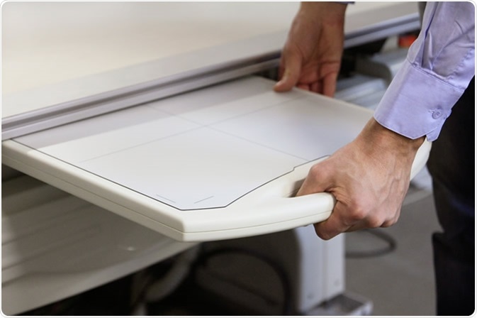
DR uses flat panel detectors based on direct or indirect conversion of X-rays to charge, which is then processed to produce a digital image.

Flat panel detector. Image Credit: Alexander Zhukovsky Design
CR uses cassette-based phosphor storage plates (PSP), which are then scanned by the computerized system into a digital format for image processing, archiving, and presentation. However, with DR, the whole procedure is digitized from X-ray detection onward.

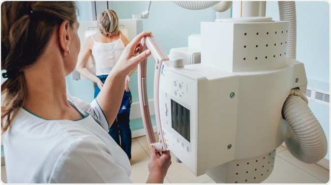
Radiologist and patient in a x-ray room. Classic ceiling-mounted x-ray system. Image Credit: Romaset / Shutterstock
Advantages of CR
- CR is the first step towards adopting digital imaging technology in many imaging centers because of the low cost required for initial installation.
- The system is compatible with most existing conventional systems, whereas DR systems come in an expensive package, and are not compatible with existing X-ray devices.
- CR can use cassettes of multiple sizes, which means the detector size can be selected to match the procedure and to increase the flexibility of positioning, whatever the area of examination.
- Portable X-ray systems can have imaging plate readers incorporated into them to provide rapid bedside radiographic examinations as well as image presentation, for speedy diagnosis.
- Single plate readers are powerful and compact and allow high patient throughput. While one plate is being processed, the next image can be acquired in rapid sequence.
Disadvantages of CR
- CR requires the cassette be removed from the X-ray machine and then placed into a reader. This is a labor-intensive step that requires the technician to leave the patient and work station with each imaging procedure, even if for a short time.
- The PSPs used in CR require longer readout and processing time.
- When single-plate readers are used, overexposures entail additional delay as the old signals are not completely erased very quickly. This means that a new plate cannot be inserted before the old plate is cleared of residual signals.
- PSP detectors are in detection position all the time. This allows them to pick up background radiation and other scattered radiation such as image noise. This is particularly important if the case if they are kept in is in or near an X-ray room.
- Another source of noise is the failure to erase signals from stored plates after more than a day or so. Taking care of multiple PSPs can entail significant labor and therefore cost, to keep inventory, clean cassettes, and assure quality.
- There is a short delay of approximately 1 minute for scanning CR plates. Cassette transfer plus plate scanning may take more time and labor than is convenient in facilities with a high patient workload, even if the machines are near each other. However, modern CR systems use plate readers that are integrated into the X-ray equipment itself, leaving little difference between CR and DR. In comparison, the DR systems house all components together and do not require any transfer of film or cassette. The time needed to produce a final DR image is 10 seconds or less.
- Image quality - PSP plates used in CR have a lower efficiency of detection compared to DR detectors. Thus, a higher radiation dose is needed to obtain adequate image resolution. However, the development of dual-side readout phosphors, as well as storage phosphors based on cesium bromide structure, has resulted in a better detection efficiency, which can match DR detectors in some cases.
Advantages of DR
- The efficiency of X-ray detection is measured by detective quantum efficiency, DQE, which is about 60%–65% with DR but only 30% with CR. Thus, the use of DR is associated with lower patient exposures because of very low imaging failure rates.
- The high speed of image acquisition is another advantage of DR technology.
- Image quality is excellent with DR whereas CR images are somewhat inferior to film X-ray systems. However, diagnostic accuracy is comparable between CR and film-based systems.
- Whereas the initial cost of CR is lower, DR systems provide high-speed workflow for technologists and speeded-up patient management, which is especially important in outpatient settings where patients need to return to home or work. Less than 1 minute is needed between exposure and image acquisition.
- Portability of DR systems is now being envisaged with the development of wireless (or previously, wired) DR detectors. This removes one of the big obstacles to the use of DR, namely, lack of portability. However, there is a long way to go before DR becomes comparable to CR in this respect. The ability to retrofit existing technology with DR detectors will mark the beginning of serious competition between CR and DR in small-scale imaging setups.
Disadvantages for DR
- The conventional DR systems are not very flexible for taking difficult views. However, newer systems are being designed to offer greater positioning flexibility.
- The initial investment costs are very high, on average up to 5 times that of a good-quality CR setup, including the reader and PSPs. This limits them to settings where return on investment is assured because of high patient throughput.
The following table summarizes the pros and cons of CR and DR:
| CR |
DR |
| Lower initial investment |
Higher initial investment |
| Can be retrofitted to existing installations |
All-new setup necessary |
| Lower image quality |
Better image quality |
| More time to final image viewing (5-7 minutes) |
Rapid image viewing (within 1 minute) |
| Labor-intensive due to the need for cassette transfer to the plate reader |
Completely digitized setup |
| Lower patient throughput |
High patient throughput |
| More bulky |
Compact profile |
| More portable |
Less portable unless newer wireless systems used |
| More flexible positioning and sizes |
Difficult to acquire awkward views |
| Higher risk of overexposure |
Lower risk of overexposure |
| Suitable for low or moderate workflow |
Ideal for high workflow |
| Less efficient |
More efficient |
| Less costly to replace |
More costly parts, requires to be protected from dropping or rough handling |
| More easy to damage and need more maintenance |
Online or remote servicing possible to cut down cost of ownership |
Further Reading
Last Updated: Nov 27, 2018