This article is based on a poster originally authored by Daniel Weekes, Blaise Louis, Sunil Modi, Erica Bello, Serena Belluschi, Tom Mitchell, Holly Herbert, Tarun Vemulkar, and Jeroen Verheyen, which was presented at ELRIG Drug Discovery 2024 in affiliation with Semarion Ltd and University of Cambridge.
This poster is being hosted on this website in its raw form, without modifications. It has not undergone peer review but has been reviewed to meet AZoNetwork's editorial quality standards. The information contained is for informational purposes only and should not be considered validated by independent peer assessment.

Cell multiplexing enables the analysis of multiple cell types in a single well, boosting data output while reducing time and reagent use. Genetic barcoding has advanced multiplexing for omic assays but not for image-based screens.
The SemaCyte® Multiplexing platform overcomes this with optical barcodes embedded in microcarriers, allowing for the identification and analysis of distinct cell lines in a single well. The Semalyse software deconvolutes these barcodes to generate sets of phenotypic images, which integrate seamlessly with open-source tools like CellProfiler and ImageJ.
This study demonstrates 4x faster results and 4x fewer reagents for compound profiling and arrayed CRISPR screens, positioning SemaCytes as a key innovation for image-based phenotypic screening.
Multiplexing cell models inside microwells using SemaCyte® microcarriers
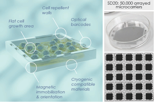
SemaCyte® microcarriers and the seeding dish 20 (SD20). Image Credit: Daniel Weekes et al., in partnership with Semarion Ltd.
In this study, SemaCyte® microcarriers were arranged as immobilized arrays on standard 20 cm2 cell culture dishes, with each dish containing 50,000 microcarriers. The microcarriers have a flat 100 x 100 μm2 cell growth area, can carry up to 30 cells, and feature cell-repellent walls embedded with optical barcodes—one unique barcode per dish.
Increased throughput
- 10x faster
- 6x more cost-effective
- Reduces plasticware
Enhanced flexibility
- Decouples culture from assays
- Reduces biological variability
Resource efficiency
- 10x fewer cells per assay
- Provides more data per cell sample
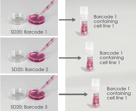
Seed cells release microcarriers into suspension and freeze inside cryovials. Image Credit: Daniel Weekes et al., in partnership with Semarion Ltd.
The cells were seeded and grown onto the SD20. Once the desired confluency and morphology were obtained, cell-containing microcarriers were released into suspension by agitating the dish. After purification, the microcarriers could be frozen as assay-ready batches inside cryovials.
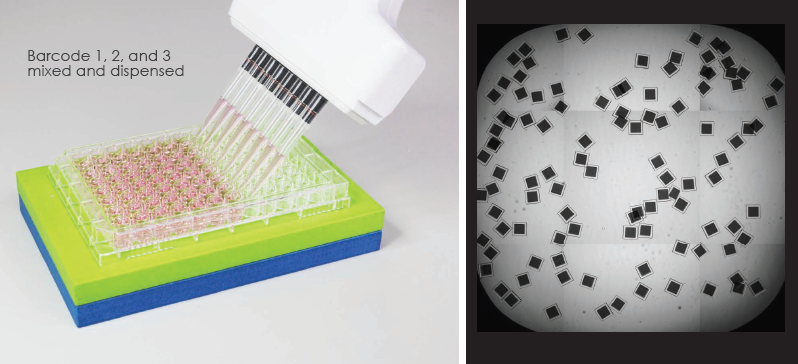
Thaw barcoded SemaCyte® microcarriers, pool various cell models, and dispense into microplates. Image Credit: Daniel Weekes et al., in partnership with Semarion Ltd.
Fresh or frozen cells on barcoded SemaCyte® microcarriers were then pooled and dispensed into microplates using standard liquid handling tools. The magnetic plate holder pulled the microcarriers to the bottom of the well and ensured they were orientated correctly.
Cryopreserved cells on SemaCyte® microcarriers were ready to assay one hour after thawing. One well of a 384-well plate could hold 250 microcarriers, enough for an 8-plex experiment, and one well of a 96-well plate can hold 1,000 microcarriers, enough for a 32-plex experiment.
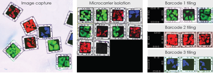
Imaging and barcode deconvolution for downstream analysis. Image Credit: Daniel Weekes et al., in partnership with Semarion Ltd.
After incubation with a compound or CRISPR library, cells on microcarriers were ready to be stained with live dyes or fixed and stained using immunocytochemistry. Images were captured with standard imaging platforms. Barcode deconvolution was carried out using the Semalyse software.
First, single microcarriers were identified and digitally isolated. Second, the optical barcodes in the brightfield channel were analyzed and matched. Third, images containing digitally tiled microcarriers were generated for each barcode or cell type. These could then be processed using typical image analysis workflows and pipelines.
Cell multiplexing for target validation & compound profiling in oncology
4-Plex cancer cell lines: Nutlin-3a sensitivity through p53 induction
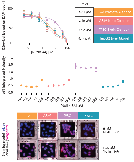
Image Credit: Daniel Weekes et al., in partnership with Semarion Ltd.
Four cancer cell lines were seeded onto barcoded SemaCytes, combined, and plated into 96-well plates for survival analysis (top) and p53 immunocytochemistry (bottom). SemaCytes were fixed at 48 hours and 16 hours post-drug addition, respectively. The data represents the mean cell count per SemaCyte, with triplicate wells and standard deviations indicated.
4-Plex Cas9 cancer cell lines: SOX2 & BCL2 as targets for oesophageal cancer
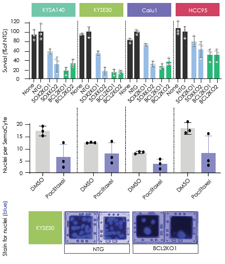
Image Credit: Daniel Weekes et al., in partnership with Semarion Ltd.
Four Cas9-expressing cancer cell lines were seeded onto barcoded SemaCytes, mixed, and plated into a 96-well plate. CRISPR editing targeted SOX2 and BCL2 and included non-targeting controls.
Cells were fixed and stained 5 days after transfection. The data shows mean cell numbers with standard deviations from triplicate wells. Paclitaxel was used as a control.
2-Plex patient-derived models: Drug 2 targets glioma, not neural stem cells
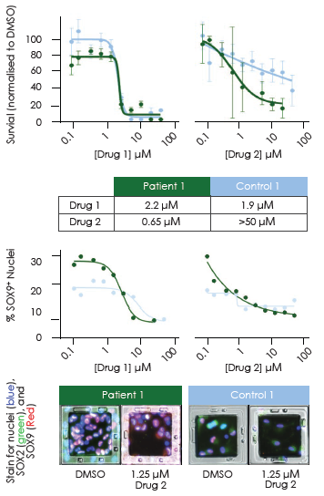
Image Credit: Daniel Weekes et al., in partnership with Semarion Ltd.
Human glioma and neural stem cells were seeded onto laminin-coated SemaCytes, mixed, and treated with two drugs. After 5 days, cells were fixed and stained for SOX2 and SOX9. Approximately five SemaCytes per cell type were analyzed for nuclear counts and SOX2/SOX9 intensity.
About Semarion
Semarion is a spin-out company from the Cavendish Laboratory at the University of Cambridge, operating at the edge of the physical and life sciences. By using microchip industry materials and techniques we are revolutionizing in vitro research on cell models to help create better medicines, faster.
Our SemaCyte® cell assaying microcarriers are flat and function as ultra-miniaturized, steerable wells to which small colonies of adherent cells are attached. We can now move and control adherent cells in liquid to accelerate, miniaturize, and multiplex cell assays. This unique approach drives 10x gains in drug screening applications such as molecular profiling, cell panel screening, or patient-derived cell work.
About ELRIG (UK) Ltd.
The European Laboratory Research & Innovation Group (ELRIG) is a leading European not-for-profit organization that exists to provide outstanding scientific content to the life science community. The foundation of the organization is based on the use and application of automation, robotics and instrumentation in life science laboratories, but over time, we have evolved to respond to the needs of biopharma by developing scientific programmes that focus on cutting-edge research areas that have the potential to revolutionize drug discovery.
Comprised of a global community of over 12,000 life science professionals, participating in our events, whether it be at one of our scientific conferences or one of our networking meetings, will enable any of our community to exchange information, within disciplines and across academic and biopharmaceutical organizations, on an open access basis, as all our events are free-of-charge to attend!
Our values
Our values are to always ensure the highest quality of content and that content will be made readily accessible to all, and that we will always be an inclusive organization, serving a diverse scientific network. In addition, ELRIG will always be a volunteer led organization, run by and for the life sciences community, on a not-for-profit basis.
Our purpose
ELRIG is a company whose purpose is to bring the life science and drug discovery communities together to learn, share, connect, innovate and collaborate, on an open access basis. We achieve this through the provision of world class conferences, networking events, webinars and digital content.
Sponsored Content Policy: News-Medical.net publishes articles and related content that may be derived from sources where we have existing commercial relationships, provided such content adds value to the core editorial ethos of News-Medical.Net which is to educate and inform site visitors interested in medical research, science, medical devices and treatments.
Last Updated: Nov 18, 2024