Enzymes in cancer diagnosis
Enzymes in cancer therapy
References
Further reading
Enzymes are proteins that facilitate biochemical reactions by acting as a catalyst, and thus, their expression and activity have a strong influence on the reactions taking place within cells. As enzymes are involved at every stage of cell regulation, a better understanding of their role and function allows them to be exploited in the clinic, both as diagnostic and therapeutic agents.

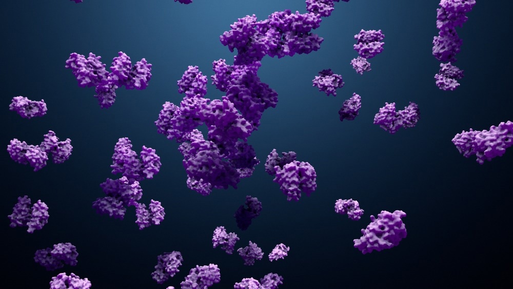
Image Credit: Design_Cells/Shutterstock.com
Enzymes in cancer diagnosis
Metabolic changes within cells are a hallmark of cancer, where genetic aberrations reduce or enhance the generation of enzymes and their metabolic products. This has allowed enzymes to be utilized as cancer biomarkers, providing a diagnostic and prognostic marker that clinicians may use to characterize a tumor.
Enzymes were among the first biomolecules utilized as cancer biomarkers, with the serine protease prostate-specific antigen (PSA) identified as a prognostic tool for prostate cancer in the 1980s. PSA is produced by the secretory cells that line the prostate glands and eventually reach the sera. Higher PSA in the blood is a result of greater numbers of more densely packed cells in the prostate, indicative of malignant cancer.
Similarly, numerous enzyme-related biomarkers for ovarian cancer have also been identified, such as the downregulation of apolipoprotein A1 and the downregulation and truncation of transthyretin.
The protein products of enzyme interactions can also be used in cancer diagnosis in the study of the proteome. The proteome consists of a large number of low and ultra-low molecular-weight protein fragments and is a rich source of information regarding the state of the cell. The proportion of specific low-molecular-weight proteins in a cell can be used to infer the activity and number of enzymes, which might otherwise be hard to distinguish from the many other proteins in the cell.
Enzymes are additionally useful in biomolecule identification and cancer diagnosis as an analytical tool in the form of the enzyme-linked immunosorbent assay (ELISA). ELISA can indicate the bonding of a target molecule colorimetrically or fluorimetrically, exploiting the highly specific binding of enzymes.
Enzymes in cancer therapy
Besides their use in cancer diagnosis, enzymes are also involved in cancer therapy, both as targets of therapeutic methods and as a therapeutic tool. For example, where an abundance of enzymes induced by dysregulated genetic transcription is causing the cancer cell to proliferate more quickly, drugs can be used to target and thus regulate the population of this enzyme, slowing the spread of cancer. Alternatively, where the absence of a particular enzyme is encouraging cancer-like behavior, introducing therapeutic enzymes can help control the proliferation of a tumor.
Administration of pro-enzymes trypsinogen and chymotrypsinogen A have demonstrated potent anti-tumor effects against a range of cell lines while limiting angiogenesis, growth, and migration. It is thought that the primary influence of these pro-enzymes is to encourage the breakdown of filamentous protein structures that grow quickly to supply the tumor with blood. Enzymes have also been used to enhance the effect of other therapeutic efforts. For example, uridine diphospho-glucuronosyltransferase (UGT) enzymes are found in the cytosol and are involved in the glucuronidation reaction, often used to remove cellular pollutants from the cytosol.
In various cancer states, upregulation of UGT is observed, which can limit the function of the anti-cancer drug irinotecan. UGT enzyme inhibitor drugs (vorinostat) have been applied to patients exhibiting high levels of UGT enzymes, demonstrating a return of efficacy of chemotherapy.
The manipulation of metabolic pathways by the inhibition of enzymes is the goal of numerous therapeutic strategies. One pathway of particular interest to cancer treatment is the management of reactive oxygen species (ROS). ROS are essential to multiple cell signaling pathways, but high concentrations of ROS can be damaging to surrounding biomolecules and induce genetic mutation and apoptosis.
Poor blood flow and acidic conditions within tumors promote the generation of ROS, and many chemo- and radiotherapeutic approaches directly induce the generation of ROS in both target and non-target cells, causing the death of cancer cells but also severe side effects. Cellular antioxidant and ROS-generating enzymes can also be damaged by the cancer therapy applied or the resulting increase in ROS concentration, further destabilizing the dynamics of ROS control within cells.
The Monoamine oxidase A (MAO-A) enzyme is involved in ROS regulation and is commonly up or downregulated in tumors. High expression of the MAO-A has been associated with poor clinical outcomes in prostate cancer patients, associated with excessive ROS concentrations. Drug inhibition of MAO-A has been found to eliminate tumor growth and metastasis in some mouse models, limiting the spread of cancer by modulating ROS generation.
References
Further Reading
Last Updated: Dec 14, 2023