Introduction
What is bladder cancer?
How can AI aid in bladder cancer diagnosis?
Using machine learning to measure tumor buds
Using AI for tumor AI detection
How can AI be used in the staging of cancer tumors?
Grading of cancer tumors using AI
Using AI to predict survival chances
Conclusion
References
Further reading
Bladder cancer (BCa) is the most prevalent urinary tract malignancy and is costly to cure. Recently, AI algorithms have been used in BCa diagnosis and outcome prediction.
These tasks include automated tumor identification, staging, and grading, bladder wall segmentation, prediction of recurrence, response to treatment, and overall survival. This article explores the significance of AI in the treatment of bladder cancer.
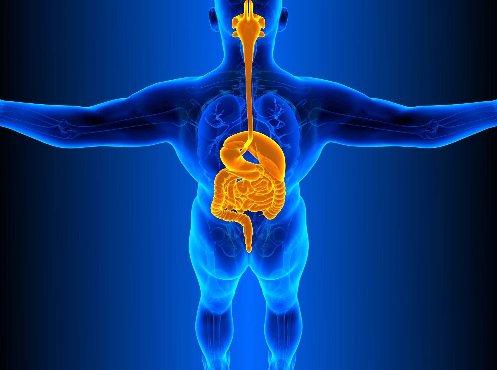
 Image Credit: Shidlovski/Shutterstock
What is bladder cancer?
Bladder cancer originates in the lining cells (urothelial cells). Urothelial cells are also in the kidneys and ureters that link the kidneys to the bladder. Bladder urothelial carcinoma is more prevalent than in the kidneys or ureters.
Non-muscle invasive bladder cancer is a heterogeneous illness whose therapy depends on muscle invasion risk. Most tumors are indolent and may be handled endoscopically. Muscle invasion can be fatal without aggressive therapy.
Various biomarkers have been examined to enhance progression prediction. Several researchers employed gene expression microarrays to find biomarkers. Array data may be interpreted using various statistical approaches, but no biomarkers have reached clinical practice.
Since statistical analysis can only be used for a large group of people and has a low success rate for predicting individual tumor behaviors, new approaches are required.
Early bladder cancer detection improves survival. Neoadjuvant chemotherapy before radical cystectomy improves survival and reduces metastatic disease. However, it can cause neutropenic fever, sepsis, mucositis, nausea, vomiting, malaise, and baldness.
It is important to examine bladder lesion response to chemotherapy to save the patient needless toxicity or to de-escalate surgery using some novel techniques.
How can AI aid in bladder cancer diagnosis?
In a multi-institutional trial, an AI-based approach enhanced clinicians' evaluations of bladder cancer patients' chemo response before radical cystectomy (bladder removal surgery).
AI can reliably detect the distribution of heterogeneous cancers using noninvasive 3D CT or MRI images (MRI). ML used with 3D MRI may discriminate low- and high-grade cancers. It allows doctors to do less invasive surgeries, minimize intra-operative blood loss, shorten hospital stays, speed gastrointestinal healing, and reduce overall problems.
 Image Credit: Magic3D/Shutterstock
Using machine learning to measure tumor buds
Researchers published an article in the journal World Journal of Urology in which they conducted a retrospective study comparing adherent and non-adherent patients in the first three years of a proposed cystoscopy regimen for demographics, histopathology, recurrence, progression, and cancer-specific mortality.
To precisely measure the tumor buds in immunofluorescence-labeled slides, the machine learning (ML) based strategy has been employed in patients with MIBC. The lymph node metastasis staging system relates to tumor bud formation. The overall survival rate of each patient with MIBC was used to place them into one of three innovative staging categories.
Using AI for Tumor AI detection
Researchers published an article in Artificial Intelligence in Medicine in which they developed a multi-layer neural network that takes the Laplacian of cystoscopy pictures (ANN) as input. The Laplacian operator draws attention to areas of the picture where the intensity values of adjacent pixels vary greatly, such as along the image's edges.
This study presents an MLP-based technique for diagnosing urinary bladder cancer. To prepare the images for this procedure, they are resized, and a Laplacian edge detector is used.
This research provides an additional option for detecting bladder cancer. This technique may be utilized as the primary or secondary algorithm in building an expert system for the diagnosis of urinary bladder cancer.
How can AI be used in the staging of cancer tumors?
The stage of a BCa tumor is defined by how deeply it has invaded the bladder. Whether the tumor has reached the detrusor muscle, patients fall into muscle-invasive (MIBC) or non-muscle-invasive BCa (NMIBC) with differing therapeutic intervention recommendations.
In an article published in Medical Physics, researchers proposed a computer-aided technique based on machine learning (ML) to detect bladder cancer stage in CT Urography (CTU). Similarly, in an article published in the Journal of Magnetic Resonance Imaging, researchers retrieved 1104 radiomics characteristics from carcinomatous ROIs in a small MRI dataset (24 NMIBC and 30 MIBC).
Researchers used recursive feature removal to winnow out non-informative features and prevent overfitting. Using pathology data, the same ML algorithms used to distinguish between NIMB and MIBC can classify phases within each group.
 Image Credit: Blue Planet Studio/Shutterstock
Grading of cancer tumors using AI
Transurethral resection of bladder tumors (TURBT) specimens must be classified reliably and reproducibly to facilitate clinical decision-making and therapy selection. In an article published in the American Journal of Pathology, researchers looked at the viability of a system that could automatically identify and grade TURBT samples.
They trained and verified two distinct neural network designs. To evaluate the digital sections, a second network is used as input for the results of the first network's urothelium identification and segmentation.
According to the consensus reading of three competent pathologists, the proposed model accurately rated 76% of the low-grade tumors and 71% of the high-grade malignancies.

 Related: Non-Invasive Diagnostic Strategies for Bladder Cancer
Using AI to predict survival chances
An accurate method of predicting how an individual cancer patient will react to chemotherapy and the patient's survival is considered a Holy grail in oncology.
For breast cancer, an accurate prediction model might help determine which individuals will have solely negative effects from chemotherapy medicines.
On the other hand, patients predicted to respond well to chemotherapy may avoid having radical cystectomy performed, greatly enhancing their quality of life.
Several researchers have used different ML methods to predict the patient's response to chemotherapy which is significantly accurate.
Conclusion
BCa is the most costly cancer to treat over the course of a patient's lifetime; hence the application of AI technology in diagnoses and prognostics can enhance patients' quality of life by reducing the number of needless radical cystectomies. Additional observational studies and new multi-phase clinical trials are needed to explore this possibility fully.
References
- Borhani, S., Borhani, R. and Kajdacsy-Balla, A. (2022) ‘Artificial intelligence: A promising frontier in bladder cancer diagnosis and outcome prediction’, Critical Reviews in Oncology/Hematology, 171, p. 103601. doi.org/10.1016/j.critrevonc.2022.103601.
- Garapati, S.S. et al. (2017) ‘Urinary bladder cancer staging in CT urography using machine learning’, Medical Physics, 44(11), pp. 5814–5823. doi.org/10.1002/mp.12510.
- Jansen, I. et al. (2020) ‘Automated Detection and Grading of Non–Muscle-Invasive Urothelial Cell Carcinoma of the Bladder’, The American Journal of Pathology, 190(7), pp. 1483–1490. doi.org/10.1016/j.ajpath.2020.03.013.
- Lorencin, I. et al. (2020) ‘Using multi-layer perceptron with Laplacian edge detector for bladder cancer diagnosis’, Artificial Intelligence in Medicine, 102(May 2019). doi.org/10.1016/j.artmed.2019.101746.
- Sun, D. et al. (2022) ‘Computerized Decision Support for Bladder Cancer Treatment Response Assessment in CT Urography: Effect on Diagnostic Accuracy in Multi-Institution Multi-Specialty Study’, Tomography, 8(2), pp. 644–656. doi.org/10.3390/tomography8020054.
- Xu, X. et al. (2019) ‘Quantitative Identification of Nonmuscle-Invasive and Muscle-Invasive Bladder Carcinomas: A Multiparametric MRI Radiomics Analysis’, Journal of Magnetic Resonance Imaging, 49(5), pp. 1489–1498. doi.org/10.1002/jmri.26327.
Further Reading
Last Updated: Dec 22, 2022