Imaging techniques used in nephrology include the following tests:
Ultrasonography
This is a non-invasive, safe and affordable imaging technique which relies on the reflections and echoes of ultrasound pusles which are directed into tissues by a transducer.
The transducer converts an electrical current into the ultrasound waves and following their reflection against the body structures, the returning ultrasound waves are converted into electrical signals read and presented by a computer as a real-time movie.

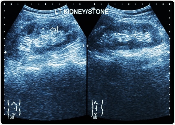
Ultrasonography of kidney : show left kidney stone ( 2 image for compare ), Image Copyright: Puwadol Jaturawutthichai / Shutterstock
Altering the ultrasound frequency can allow investigation of the kidney and bladder even during pregnancy.
It can be useful for the following:
- Acute kidney injury e.g. post-renal obstruction.
- Chronic kidney disease (sometimes indicated by decreased kidney size)
- Congenital anomalies - eg, hypoplasia, agenesis, duplex systems.
- Detection of renal cysts, abscesses and neoplasms e.g. simple cysts, polycystic kidneys.
Nuclear medicine
This encompasses techniques such as nuclear medicine renal scans. This type of scan involves imaging to study efficiency of each kidney’s function by considering the blood supply, function and excretion of urine from each kidney.
The scan can be performed with 2 different substances - diethylenetriaminepentaacetic acid (DTPA) or mercaptoacetyltriglycine (MAG3).
The former is often more commonly used and both are similar, but MAG3 gives significantly better images in some patients (e.g. very young children and patients with poor kidney function).
A DTPA Scan may also be undertaken to evaluate:
- Function and perfusion of renal tubules and transplants
- Renovascular hypertension
- Condition of renal tubules due to obstruction, trauma or damage
- Renal artery stenosis
Computed Tomography (CT) scan
CT scans are not routinely used to check for problems – normally, the onset of symptoms might call for it. These scans involve taking a series of X-rays of the patient’s body at slightly different angles. Then this collection of X-ray images is interpreted and superimposed by a computer and displayed as a 2-dimensional form.
CT scans may be done with or without "contrast" in which contrast is a substance ingested or injected into an intravenous (IV) line in order to allow clearer visualisation of the particular organ or tissue under study. If contrast media is used, there is a risk of an allergic reaction, therefore, patients exhibiting allergies or sensitivity to medications or who have had prior reaction to any contrast media, should inform their doctor. A disadvantage of CT is the exposure to significant amounts of ionizing radiation.
CT scans provide more detailed imagery than standard kidney, ureter and bladder (KUB) X-rays. CT scans of the kidneys are used to detect conditions including tumors or other lesions, obstructive conditions (e.g. kidney stones), congenital anomalies, fluid accumulation surrounding the kidneys, and the location of abscesses.
CT Kidneys and Bladder - Five pathologic cases discussed
Magnetic Resonance Imaging (MRI) scan
MRI is an alternative to the CT scan that can be used in all patients except those with pacemakers due to the harmful effects of strong magnetic fields.
MRI imaging is based on the behavioural change of hydrogen nuclei (i.e. protons) when subjected to a magnetic field (due to the MRI system). The nuclei act as miniature magnets, which can be manipulated in an MRI image in order to distinguish among various tissue characteristics.
MRI has emerged as an attractive approach due to its non-invasiveness and suitability for repeated application. Because of this, they are used to monitor the functioning of renal allografts and accurately characterize renal tumor aggressiveness in order to guide management.
Its advantages over CT scanning include its greater soft tissue contrast and increased safety due to the avoidance of ionizing radiation and iodinated contrast compounds. Most importantly, combinatorial techniques exist to probe different aspects of a tumor such as microstructure, vascularity, and oxygenation including dynamic contrast enhanced (DCE), (diffusion weighted imaging) DWI and blood oxygen level dependent (BOLD) MRI.
References
Further Reading
Last Updated: Dec 30, 2022