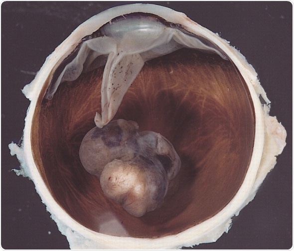
Choroidal melanoma is among the most common tumors of the eye. It affects one of the most vascular tissues of the body. Its diagnosis requires an internal ophthalmoscopy as well as ancillary tests including fluorescein angiography, ultrasonography of the eye, and transillumination.
Fine needle aspiration cytology has now fallen out of favor due to the risk of tumor seeding in the needle track, as well as the low reliability of negative cytology results. The condition is one with a high risk of metastasis and is difficult to treat adequately.
The treatment of choroidal melanoma has several forms.
These include:
- Visual acuity in the affected and the normal eye.
- Tumor characteristics, including its size and situation, the involvement of other ocular tissues, and the presence of distant metastasis.

Variably pigmented, mushroom-shaped choroidal tumor has ruptured the Bruch membrane and grown into the subretinal space. Image Credit: The Armed Forces Institute of Pathology (AFIP) - PEIR Digital Library (Pathology image database). Image# 406734.
Small Melanomas of the Choroid
When the tumor is small, it may be very difficult to distinguish it from a choroidal nevus. Some features suspicious of malignancy include:
- The tumor is over 2 mm thick.
- Presence of fluid.
- Subretinal fluid accumulation.
- Orange lipofuscin pigment at the retinal pigment epithelial surface.
- Tumor margin extends anteriorly to touch the optic disc.
- Hollow appearance on ultrasonography with acoustically silent zones and shadowing associated with vascular pulsations.
- The absence of halo and of drusen.
Where such features are absent, the patients may be followed up with a detailed checkup at 6-monthly intervals, for at least one year, followed by yearly examinations thereafter provided the condition shows no change. If one or two features suggest malignant change, the patient should be called in every 4-6 months instead for review.
If malignant changes are present, the clinician may choose from several options depending on the findings:
- Laser photocoagulation.
- Photodynamic therapy.
- Plaque radiotherapy.
- External beam radiotherapy.
- Transpupillary thermotherapy – very limited in scope by the laser’s inability to penetrate thicker layers of tumor, and so associated with higher rates of local recurrence.
- Local resection of the tumor.
- Enucleation of the eye.
For Medium-Sized to Larger Melanomas
Enucleation
When a choroidal melanoma is associated with severe secondary glaucoma precipitated by several tumor-related factors, or with invasion of the optic nerve, enucleation of the eye was formerly done. Currently it is often preceded by external beam radiotherapy with helium ions or protons - this is to address the concerns of metastasis precipitated by enucleation. However, enucleation either alone, or with preoperative radiotherapy, is associated with comparable long-term survival rates. It is indicated with diffuse tumor spread or evidence of extraocular tissue involvement.
Plaque Brachytherapy
In other cases, plaque radiotherapy is used to spare the eye. A plaque containing seeds of radioactive iodine-125 (which is used most frequently), gold-198, palladium-103 or other similar metals is sutured to the sclera to deliver local radioactive energy and destroy the tumor cells. This is possible only if a 2mm base is available around the tumor margin, allowing the delivery of adequate energy to the entire tumor. Exact assessments of the tumor base diameter depend upon techniques such as transillumination, ultrasonography or the use of optic disc diameter. The plaque is confirmed to be in position by ultrasound, ophthalmoscopic examination or the use of MRI.
The plaque has to remain in place for 3-7 days, the interval varying with the tumor size and the rate at which the radiation enters the tumor. For instance, with I-125, 80-100 Gy is the recommended dose to the tumor apex, delivered at 5-125 cGy/hour.
Even with local radiotherapy, however, later complications may set in, or the tumor may recur, necessitating eventual enucleation. The adverse effects of radiotherapy include secondary neovascular glaucoma, retinopathy, the onset of cataractous changes, or vitreous hemorrhage. Recurrence occurs in 12 of 100 patients and is in most cases near the original tumor site. Thus persistence of tumor cells, deterioration of vision in the treated eye, and chronic irritation of the eye are potentially worrisome outcomes of radiation. The cost difference between enucleation and radiation with plaque insertion is also significant. It is noteworthy that in both enucleation and brachytherapy, visual acuity declines after 3-5 years.
External Beam Irradiation
External beam irradiation has also been used to treat choroidal melanomas. It is claimed that delivery of radiation is better spread at optimal dosage over the whole tumor, in addition to greater sparing of surrounding tumors, increased accuracy and the ability to treat larger tumors more successfully. This technique uses helium nuclei or protons, both charged particles, and is estimated to cost twice as much as an enucleation surgery. However, quality of life is comparable with both procedures. Maculopathy occurs in almost 2 of every 3 patients with tumors within 4 disc diameters of the macula following external beam irradiation.
Gamma Knife
A third option is the use of the gamma knife, or stereotactic radiation surgery, which is another radiation procedure with greater precision and cost.
Sclerouvectomy
Surgical treatment avenues include enucleation as well as conservative procedures. One of these is sclerouvectomy, which includes tumor removal with part of the sclera and retina. The scleral gap is closed using banked scleral tissue. A more tissue-sparing procedure is lamellar sclerouvectomy, which attempts to prevent vitreous and retinal complications.
Orbital Exenteration
Orbital exenteration includes removal of the entire contents of the orbit, a procedure adopted when there is extensive spread of the tumor within the orbit. However, this should be considered a palliative procedure in view of its disfiguring nature and poor prognosis for survival, given the widespread nature of the disease.
Choosing a Procedure
Overall, it is thought advisable to use as conservative approaches as are compatible with reducing tumor-associated risk of fatality and metastasis. It is already known that micrometastases are present in most patients with choroidal melanoma at the time of diagnosis.
When the tumor presents with distant metastasis, systemic chemotherapy is the treatment of choice, with local treatment being palliative in nature. With all treatments for this tumor, the concerns of the patient must be taken into account, considering that two serious losses are in view – the loss of vision or of the eye itself, against a possible loss of life.
References
Further Reading
Last Updated: Aug 21, 2023