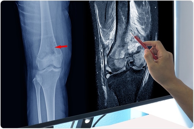
The symptoms of osteosarcoma may not be obvious in the beginning and, as a result, the initial diagnosis of the disease often occurs after it has already developed significantly. In fact, the first time that many individuals notice a sign of the disease is when they suddenly fracture the affected after minor trauma as a result of the weakened bone.

Image Credit: April stock / Shutterstock.com
There are, however, some signs that may be evident. Therefore, if it is possible to recognize these signs earlier in the course of osteosarcoma, timely treatment can be initiated earlier to improve the patient's prognosis and avoid fractures.
When the disease is suspected, several imaging techniques may be used to investigate the structure of the bone and identify whether there is a tumor present or not. A biopsy of the tumor is then needed to confirm the malignancy of the tumor and the appropriate treatment.
Presenting symptoms
Pain is a symptom of osteosarcoma that many patients notice. This is particularly true in teenagers who participate in sporting activities as these individuals tend to complain about pain below the knee or in their lower femur. It is common for this pain to worsen during the nighttime. Additionally, it is normal for the pain associated with osteosarcoma to vary in intensity and come and go over time.
Swelling in the area may also become evident, especially if the tumor has grown to a large size. However, even a big tumor may not produce visible swelling if it is not near the surface of the body. An example of this is a tumor on the pelvis, which is more difficult to detect.
The bones affected by osteosarcoma are not as strong as healthy bones. This is not often visible or identifiable by patients; however, they will be more likely to fracture the bone as a result of this. These fractures of weakened bones can occur even with minor trauma, which is also known as a pathological fracture.
Diagnostic imaging
Most tumors of the bone are benign; however, adequate imaging needs to be done to accurately diagnose the condition.
An X-ray is the first type of imaging that needs to be done, which is then followed by a number of different scans. This includes:
- Computed tomography (CT) scan
- Positron emission tomography (PET) scan
- Bone scan
- Magnetic resonance imaging (MRI)
It is characteristic to see Codman’s triangle in the X-ray image, which is a subperiosteal lesion formed when the tumor causes the periosteum to rise. Additionally, it is possible for the tumor to metastasize to the lungs, which will appear on the X-ray image. This metastasis of osteosarcoma will usually present as a nodule on the lower part of the lung.
Osteosarcoma in Children: Jude's Story | WebMD
Tumor biopsy
The final step to confirm the diagnosis of osteosarcoma is to take a surgical biopsy. This enables physicians to test the tumor cells for malignancy and help guide future treatment approaches.
Diagnostic images and scans are an effective way to understand the nature of the tumor. However, the only definitive way to determine if the tumor is malignant or benign is to take a bone biopsy.
A qualified orthopedic oncologist should conduct this procedure, as it is of utmost importance that is carried out correctly. If it is not performed properly, it may be difficult to salvage the affected limb and avoid amputation.
Categorization
There are several different variants of osteosarcoma. Once a diagnosis has been confirmed, the disease is usually categorized as:
- Conventional (e.g., osteoblastic, chondroblastic, or fibroblastic)
- Telangiectatic
- Small cell
- Low-grade central
- Periosteal
- Paraosteal
- Secondary
- High-grade surface
- Extraskeletal osteosarcoma describes the types of cells affected and can help with the treatment decisions of the condition.
Further Reading
Last Updated: May 22, 2021