When patients present with pain in the ear, which is one of the symptoms of otitis media, it is important to make an accurate diagnosis using appropriate techniques, as the pain may be indicative of another condition.
Clinical examination
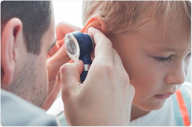
Clinical history alone is not sufficient to determine the involvement of otitis media. Therefore, visualization of the tympanic membrane is needed to make a diagnosis. This technique is usually conducted with a pneumatic otoscope attached to a rubber bulb, which helps to view the tympanic membrane and assess its mobility.

Image Credit: Mala Iryna / Shutterstock.com
The signs that are indicative of otitis media upon visual inspection of the membrane include:
- Bulging and fullness
- Cloudiness
- Redness (erythema)
Occasionally, it can be difficult to confirm the diagnosis with solely a visual examination of the eardrum. This may happen because the ear canal is very small, which makes it difficult to get a clear view. Earwax can also obstruct the view through the ear canal. In the event that earwax is preventing the view, it may be removed with a blunt cerumen curette or wire loop.
It is also possible to make a false diagnosis based on the circumstances when the diagnosis was made. For example, if a young child is upset and crying, the eardrum may look red and inflamed and thus appear similar to otitis media as a result of the distension of small blood vessels on it.
Differential diagnosis
The two most common types of otitis media inckude acute otitis media (AOM) and otitis media with effusion (OME). It is important to differentiate between these during diagnosis, as the treatment differs significantly, particularly in regards to the use of antibiotics. It has been suggested by some practitioners that the best way to determine the type of otitis media is by observing the bulging of the tympanic membrane.
Acute otitis media (AOM)
For children that exhibit moderate to severe inflammation of the tympanic membrane or have recently noticed otorrhea, which is the term used to describe drainage from the ear, the symptoms are not likely to be due to external otitis. In addition, mild bulging of the eardrum with pain onset within 48 hours and intense redness is diagnostically indicative of AOM.
Otitis media with effusion (OME)
OME can also be referred to as serous otitis media or secretory otitis media. OME is a build up of fluid or effusion that occurs with the middle ear as a consequence of Eustachian tube dysfunction and the resulting negative pressure in the middle-ear space.
There may not be any pain or bacteria infection associated with OME; however, the fluid within the middle ear can inhibit hearing ability when it interferes with the normal sound wave vibration of the eardrum.
Over the timespan of several weeks, the fluid in the middle ear can become very thick and resemble the consistency of glue, which has led to this condition being referred to as glue ear occasionally. This also increases the likelihood of associated conductive hearing impairment.
Some risk factors for OME within the first two years of life are:
- Feeding infants whilst lying down
- Child care from an early age
- Smoking by parents
- Minimal or lack of breastfeeding
- Large amount of time in childcare
Other types
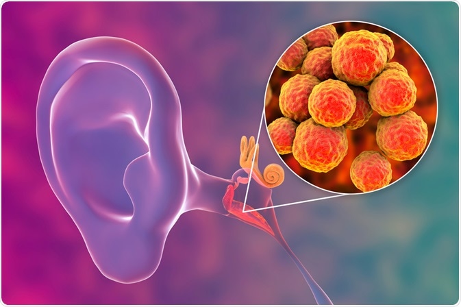
Chronis suppurative otitis media involves a bacterial infection of the middle-ear that persists for several weeks or longer and is accompanied by a hole in the tympanic membrane. The pus may accumulate and drain outside the ear, although it may also only be visible on inspection with an otoscope or binocular microscope. This condition is more common in individuals with poor Eustachian tube function and often precipitates hearing impairment.

Image Credit: Kateryna Kon / Shutterstock.com
Viral otitis may present with blisters on the outside of the tympanic membrane, which is also known as bullous myringitis. Another type of otitis media is adhesive otitis media, which involves a thin retracted eardrum that is vacuumed into the middle-ear space and sticks to the ossicles and bones in the middle ear.
References
Further Reading
Last Updated: Sep 24, 2022