Initially described as “plasma cell pneumonia”, pulmonary infection with the microorganism Pneumocystis jiroveci (formerly known as Pnemocystis carinii) was first identified in malnourished infants, especially those in orphanages, at the conclusion of the Second World War. Today this disease, manifesting as a severe and often fatal pneumonia in immunocompromised patients, is of increasing importance to pediatricians and other specialists around the world.

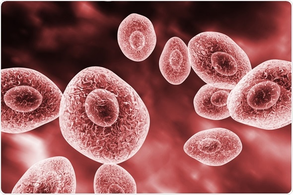
Pneumocystis jirovecii, opportunistic fungus which causes pneumonia in patients with HIV, 3D illustration - Image Copyright: Kateryna Kon / Shutterstock
Pneumocystis jiroveci is an opportunistic unicellular fungal pathogen that is widespread in the environment and is thought to enter the body via airborne transmission. It also represents one of the several microorganisms that can cause life-threatening infection in patients with advanced HIV infection and AIDS.
Pneumocystis Pneumonia (PCP): Part I
Epidemiology of Pneumocystis pneumonia
In the 1960s, Pneumocystis pneumonia was observed predominantly in children who had specific congenital defects of the immune system, as well as in children and adults with acquired immune defects due to malignancy or its treatment. When organ transplantation became more widespread in clinical medicine, it was found that this type of pneumonia was also linked to the immunosuppressive regimens used to prevent organ rejection.
In the 1980s, Pneumocystis pneumonia was found to cluster in some previously healthy men. This subsequently prompted a search for the underlying cause of immunosuppression in these patients, which eventually led to the first definition of the acquired immune deficiency syndrome (AIDS). And indeed, it turned out that a majority of individuals who develop Pneumocystis pneumonia have abnormal function and/or number of T lymphocytes.
Today, this type of pneumonia is still the ubiquitous AIDS-defining diagnosis in North America, Europe and Asia, albeit it is mostly restricted to patients who are unaware of their HIV status at presentation, as well as to those who are intolerant of or non-compliant with antiretroviral therapy.
Before adequate prophylaxis was introduced, attack rates for Pneumocystis pneumonia ranged from 25% in individuals with rhabdomyosarcoma (one of the most common types of soft tissue cancers) to 43% in those with acute lymphoblastic leukemia (a fast-growing cancer that affects a certain type of white blood cells).
Transmission and colonization
A majority of healthy children and adults have antibodies to Pneumocystis jiroveci. This finding prompted the initial hypothesis that Pneumocystis pneumonia could arise in an immunosuppressed patient due to reactivation of a childhood-acquired, latent infection. However, additional genotypic analyses have revealed that the primary infection can be effectively cleaned out by the immunocompetent host. This shows that this type of pneumonia is the result of re-infection with different strains rather than reactivation of a pre-existing infection.
The transmission of Pneumocystis jiroveci cysts is airborne, but its presence in the lungs is usually asymptomatic. Nevertheless, individuals with impaired immunity, and most notably those with a low CD4+ T cell count (below 200 per μl) are considered at risk for the development of Pneumocystis pneumonia. These cells play a pivotal role in mediating the adaptive immune response to a plethora of pathogens.
Research shows that immunocompetent hosts are potential reservoirs of the fungus in the population, as they can transfer it from one person to another over short distances. Animal studies have demonstrated that even close-contact periods as short as one day can result in transferring Pneumocystis jiroveci from donors to immunocompetent recipients.
Other than immunosuppression, important risk factors for Pneumocystis colonization and Pneumocystis pneumonia include pulmonary disorders and infection with cytomegalovirus (CMV). The latter is due to the suppression of both helper T cells and antigen presenting cells by the virus, which alters the host immune response.
References
Further Reading
Last Updated: Feb 27, 2019