The importance of studying protein-protein interactions
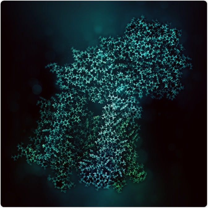
Proteins do not always function alone, and protein-protein interactions are thought to be important in cellular processes. Most of them will interact with other proteins as oligomers; this could be between two of the same proteins (homo-oligomer) or between different proteins (hetero-oligomer).
Within a bacterial genome, it is estimated that up to 80% of cellular proteins function as an oligomer, with homo-oligomers being four times more common than hetero-oligomers.
However, the definition of “oligomer” can vary according to the techniques used, and how the protein functions – i.e. does it function alone or with other proteins?

Image Credit: Studio Molekuul/Shutterstock.com
How do you visualize protein-protein interactions using fluorescence?
Various methods can be used to study protein oligomerization.
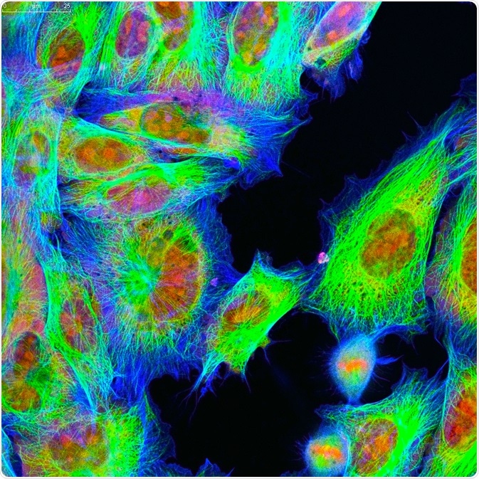
One such method involves tagging proteins with a fluorescent label so that it can be seen under a fluorescent microscope.
This can be genetically coded, organic dyes and nanoparticles such as quantum dots. Factors which need to be considered include the brightness and the “quantum yield” (the probability of a photon being emitted when the fluorophore is excited) of the fluorophore, which are linked to the environmental conditions, such as pH and viscosity, as well as the structural property of the fluorophore such as rigidity.
The strategy which is most commonly used is genetically coded fluorescent tags. This involves adding the nucleotide sequence of a fluorescent protein, such as Green fluorescent protein (GFP) and red fluorescent protein (DsRed), on to an end of the coding sequence for the protein of interest.
To ensure that the addition of the fluorescent tag does not affect the function of the protein, it is important to consider the most appropriate end to add the fluorescent tag, as well as using an appropriate linker between the protein and the fluorescent tag.
Organic dyes can be added directly or indirectly to the protein, and have advantages including stability and smaller size compared to fluorescent proteins. However, unlike genetically coded tags, these can lead to high levels of background noise due to the presence of unbound dyes.
The method which is commonly used to add organic dyes involves covalent modification of amino acid chains, as well as the use of fluorescently labeled immunoglobulins (immunofluorescence).
Quantum dots are semiconductor crystal nanoparticles and these have gained attention as a versatile and sturdy fluorescent label. The main advantages of using quantum dots include the ability to tune the emission spectra, and increased quantum yield and photosensitivity compared to organic dyes. However, these nanoparticles are generally thought to be too large to be able to diffuse through the cell membrane.
Examples of fluorescence microscopy-based methods
Once proteins are tagged with fluorescence, several methods can be used to study them using fluorescence microscopy-based methods; examples include the use of bimolecular fluorescence complementation and quantitative single-molecule localization microscopy.

Image Credit: Vshivkova/Shutterstock.com
Bimolecular fluorescence complementation has been developed to facilitate the study of protein-protein interactions.
This can be possible in solutions or living cells, and was developed after the observation that fluorescent proteins can be coded as separate components; each part of the oligomer would carry the component of the fluorescent protein, and once they connect the components of the fluorescent protein will combine and thus the fluorescent protein can only emit fluorescence when the protein is oligomerized. This then makes it possible for protein oligomerization to be visualized under a fluorescent microscope.
One such example is the use of quantitative single-molecule localization microscopy. Single-molecule localization microscopy has made it possible to investigate the stoichiometry of protein complexes in situ. This includes subunit counting, which would enable protein oligomerization to be studied.
Here, photobleaching of a fluorophore is monitored by using a total internal reflection fluorescence (TIRF) microscope set-up. Fluorescence is kept low, and the TIRF microscope is set so that only the membrane proteins are illuminated.
This helps to eliminate background fluorescence as well as fluorescence coming from the cytoplasm. It is then possible to effectively isolate individual subunits which are emitting fluorescence. Thus, it is possible to go beyond high-resolution imaging and count individually tagged molecules such as a protein or a protein subunit.
A study has used single-molecule localization microscopy to study the oligomerization of human glycine receptor.
Source
- Petazzi, R. A. et al. (2020) Fluorescence microscopy methods for the study of protein oligomerization. Chapter 1, Progress in Molecular Biology and Translational Science volume 169 https://doi.org/10.1016/bs.pmbts.2019.12.001
Last Updated: Apr 15, 2020