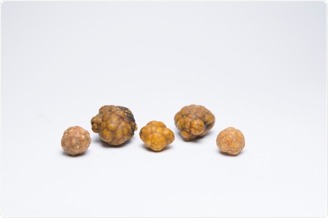
Bladder stones are hard masses of mineral that form in the bladder when it does not empty entirely.

Image Credit: harnchoke punya / Shutterstock.com
Some patients with bladder stones may not notice any symptoms, as the lumps are small enough to pass out of the bladder and can be excreted in the urine. However, other patients with larger stones that irritate the bladder wall or obstruct the urine flow report several symptoms.
Diagnosis of bladder stones involves an initial consultation to discuss medical history and a physical examination. This is often followed by diagnostic imaging techniques, such as a spiral computed tomography (CT) scan, X-ray, or ultrasound to confirm the presence of bladder stones.
Symptoms
Symptoms of bladder stones are usually only evident when the stones irritate the bladder lining or obstruct the flow of urine. Some of the common symptoms that a patient presenting with bladder stones may complain of include:
- Moderate to severe lower abdominal pain
- Difficulty or pain upon urination
- Frequent urge to urinate
- Cloudy or dark urine
- Hematuria
- Pain in or around penis (in men)
It is common for the flow of urine to suddenly stop and the patient to experience some pain in the penis, scrotum, perineum back or hip. Additionally, many patients with bladder stones also have a urinary tract infection (UTI), with symptoms such as pain and fever. This is because a UTI can both cause and result from bladder stones.
Initial diagnostic tests
The diagnosis of bladder stones usually begins with a comprehensive medical history, including past procedures in the pelvic region.
A physical exam is the next step to investigate the cause of the symptoms and specifically investigate whether there is any enlargement of the bladder in the lower abdominal region. A rectal examination is also usually needed for men that present with symptoms, as it can reveal an enlarged prostate or other related problems.
A urine culture is often collected for urinalysis to identify whether blood, bacteria and/or crystallized minerals are present within the urine. A urine culture is also useful in identifying a UTI.
In some cases, cystoscopy may also be indicated to further investigate the cause of the symptoms and make an accurate diagnosis.
What Are Bladder Stones?
Diagnostic imaging techniques
The most common imaging technique used in the diagnosis of bladder stones is a CT scan, which creates cross-sectional images of the abdominal region to detect bladder stones. A spiral CT scan is a more efficient and rapid technique that provides greater definition of the internal structures and can also detect even small stones present within the bladder.
An intravenous pyelogram is a special type of imaging test that involves the injection of a contrast material into a vein that is excreted through the renal tract. X-ray imaging is then used at regular intervals throughout this process to observe the movement of the material and check for bladder stones.
Ultrasound uses the reflection of sound waves off the organs and internal structures to visualize the bladder and detect any stones present.
X-ray imaging can sometimes be used on its own to detect the presence of bladder stones in the urinary tract; however, this test is less sensitive and does not show all stones.
It is important for patients with bladder stones to be diagnosed as soon as possible to ensure that they receive adequate treatment for the condition and minimize any complications that may occur.
References
Further Reading
Last Updated: Mar 18, 2021