The retinoid cycle, also called the visual cycle, is a complex system which replenishes the compounds that are needed for light to activate the receptors in the eye. This cycle was first described in detail by George Wald, for which he won the Nobel Prize in Physiology or Medicine in 1967.
Anatomy and Physiology
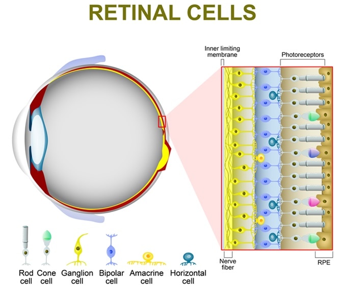
There are three important photoreceptors found in the retina in the back of the eye. The main two types are the rod cells and the cone cells. Rod cells are involved in low light vision and are found around the edges of the retina, therefore they are also important in peripheral vision.

Rod and cone cells. The arrangement of retinal cells is shown in a cross section. Image Credit: Designua / Shutterstock
Cone cells are mainly found in the center of the retina and mediate the detection of different colors. There are 3 types of cone cell, which are activated by different wavelengths of visible light. When a certain combination of these 3 types of cone cell are activated, this equates to a particular color that the brain can interpret.
Both rod cells and cone cells contain a type of protein called opsins, which are light-sensitive and are needed for phototransduction – where light hitting the retina is turned into an electrical signal via the optic nerve to the brain. Opsins are G-protein-coupled receptors, so when activated, they activate a cascade of other reactions which leads to the production of electrical signals to the brain.
Opsin molecules combine with chromophore 11-cis-retinal to form rhodopsin, which is the main pigment found in rod cells. 11-cis-retinal is converted to all-trans-retinal when light stimulates the photoreceptors, and it is released from opsin. In order for the cells to be stimulated again, and for more signals sent to the brain, all-trans-retinal needs to be recycled back into 11-cis-retinal.
Most of this recycling occurs in a section of the retina called the retinal pigment epithelium, or RPE cells. The RPE is a layer of cells located in the outermost layer of the retina, and attached to the choroid, where blood vessels are located.
Retinoid Cycle Steps
The cycle begins after light hits the retina and stimulates a photoreceptor, producing all-trans-retinal.
- All-trans-retinal is pumped out of the photoreceptors by the transporter ABCA4, and reduced into all-trans-retinol by retinol dehydrogenases RDH8 and RDH12.
- All-trans-retinol diffuses into the RPE cells by binding to interphotoreceptor retinoid-binding protein (IRBP).
- Lecithin retinol acyltransferase (LCAT) esterifies all-trans-retinol into retinyl esters.
- These retinyl esters are stored in retinosomes which are lipid bodies in the RPE cells used for storage of metabolites.
- The retinyl esters can then be modified again by RPE65 into 11-cis-retinol
- Finally, RDH5 oxidises 11-cis-retinol into 11-cis-retinal.
- 11-cis-retinal then diffuses back out of the RPE cell into the photoreceptor with the help of IRBP, where it recombines with opsin ready for stimulation.
Associated Diseases
This cycle is very important in allowing continuous phototransduction in the retina. Without this cycle, the eye would not be able to function.
With many of the enzymes and other proteins involved in the retinoid cycle, there is a possibility of changes (or mutations) that can cause a variety of diseases. For example, a mutation in ABCA4 or an RDH can result in age-related macular degeneration (AMD), or Leber congenital amaurosis (LCA) which are both diseases which cause a loss of vision. Other possible diseases include congenital stationary night blindness (CSNB) and retinitis pigmentosa.
11-cis-retinal and all-trans-retinal are types of vitamin A. This means that the retinoid cycle can be affected by vitamin A deficiency. This is a serious problem, especially in children, with an estimated 250 million preschool children affected worldwide.
Further Reading
Last Updated: Apr 20, 2022