Understanding thoracic imaging
Early detection through imaging
Differential diagnosis made clear
Monitoring disease progression and treatment response
Future directions in thoracic imaging
Conclusion
References
Further reading
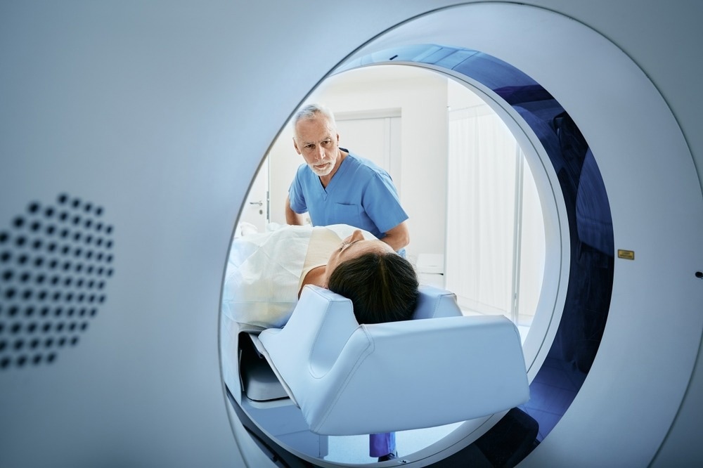
Thoracic imaging is critical in detecting, diagnosing, and treating diseases related to the lungs and surrounding organs in the chest. Advanced imaging techniques provide vital insight into lung health and various respiratory conditions in patients to accurately deliver effective treatment plans.

Image Credit: Peakstock/Shutterstock.com
Understanding thoracic imaging
Modalities available for thoracic imaging include chest X-rays, computed tomography (CT), MRI, and imaging related to nuclear medicine, such as ventilation-perfusion lung scanning and positron emission tomography (PET).
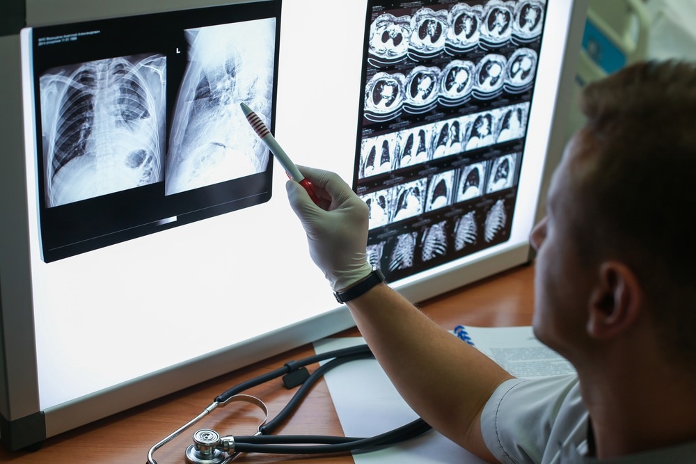
Chest X-rays are usually the starting point for patients with potential respiratory problems. They use a focused beam of radiation to show posterior-anterior and lateral viewpoints that provide an overview of the lungs as well as the cardiovascular system. A few common indications in patients that may result in healthcare professionals requesting an X-ray may include, but are not limited to, an infection, major trauma, acute chest pain, asthma, bronchiolitis, or a suspected mass.
CT scans are typically the second step after a chest X-ray has revealed an abnormality that may require further evaluation or may be used when there is a prolonged disease course or if another illness or condition is suspected. Common indications that may cause a doctor to request a CT scan include a pulmonary mass that may have been identified in an X-ray, or if a malignant disease is suspected, such as lymphadenopathy and metastatic disease detection.
MRI scans use magnetism and radio waves to take pictures of the inside of the body and provide improved soft tissue contrast compared to CT scans. This type of scan can be used to find a tumor and stage of cancer and is usually used for assessing congenital cardiac and vascular diseases.
Additionally, a PET/CT scan is considered a powerful molecular imaging technique for staging primary lung cancer, where there may be a moderate-to-high risk of malignancy.
Early detection through imaging
Thoracic imaging is vital for the early detection of respiratory diseases in patients with suspected diseases and underlying malignancies, such as pneumonia and lung cancer.
Healthcare professionals can use chest X-rays to diagnose or treat conditions such as pneumonia, emphysema, or chronic obstructive pulmonary disease (COPD). Additionally, low-dose CT scans are currently the standard of care imaging technique used to screen patients at high risk for lung cancer.
Early disease detection is highly important for patient prognosis, with better treatment outcomes and improved quality of life. For example, early identification of lung cancer can increase the range of treatment options available, such as being suitable for targeted therapies based on specific gene mutations, which can greatly impact patient survival rates.
Differential diagnosis made clear
Critical use of various thoracic imaging includes the ability to differentiate between a range of respiratory conditions that share similar symptoms. Similar symptoms of respiratory diseases encompass shortness of breath, persistent or severe cough, wheezing, and difficulty breathing, to name a few. These symptoms can belong to a range of diseases, from COPD to influenza or COVID-19 and even lung cancer.

Image Credit: Egor_Kulinich/Shutterstock.com
The use of thoracic imaging can help inform healthcare professionals which respiratory disease these symptoms belong to, along with other tests, including a spirometry test, which assesses how well a patient’s lungs are working, and blood tests.
Once an accurate diagnosis is reached, healthcare professionals can develop appropriate and effective treatment plans for a patient. For COPD, there is currently no cure. However, treatments can be used to slow the progression of the condition and control the symptoms.
For non-small cell lung cancer (NSCLC), there may be an opportunity for a patient to receive targeted therapy or immunotherapy, depending on if the patient meets the criteria for treatment.
Monitoring disease progression and treatment response
Interestingly, thoracic imaging can be used for non-diagnostic applications, including monitoring disease progression and assessing a treatment's efficacy. An example of this includes lung cancer, which requires patients to receive follow-up care with chest CT scans to monitor if the cancer has been fully eliminated and check for recurrence. The risk of recurrence is due to a small number of cancer cells that may have remained undetected in the body, causing cancer regrowth over time.
Patients with NSCLC are at a higher risk of developing a second lung cancer, which is why future scans are recommended to monitor for either cancer recurrence or the possible development of a new lung cancer.
The American Society of Clinical Oncology (ASCO) recommends that most patients successfully treated for stage I to stage III NSCLC should receive imaging every six months for the first two years after their treatment to monitor for cancer recurrence. After this period, patients are recommended to receive a low-dose chest CT scan once a year.
Future directions in thoracic imaging
Artificial intelligence (AI) is considered to hold great potential as an analytic tool for thoracic imaging with a wide spectrum of possible applications, such as reducing image noise as well as radiation dose. AI has shown a high level of performance in interpreting images within medicine. However, how these tools can help physicians within clinical practice is not yet established.
Pneumonia and respiratory symptoms are among the most frequent reasons for emergency room visits in the US, with radiologic studies being a common primary evaluation tool – this has only increased over the past two decades.
With the increasing demand for imaging tests and the lack of physicians and radiologists to complement this requirement, AI-assisted analysis can directly improve the clinical outcomes of patients with diseases that need timely diagnoses.
A research study that performed simulated reading tests for chest X-rays from emergency room patients with and without AI-assistance found that radiologists detected significantly more critical and urgent abnormalities with the use of AI-assistance. AI-assistance was also shown to reduce the time period of reporting chest X-rays of critical and urgent cases.
Conclusion
Thoracic imaging plays a significant role in respiratory diseases, with advanced imaging technologies used to detect diseases, including cancer. Imaging is also critical for accurate diagnoses of diseases with similar symptoms, leading to effective treatment as well as management of cancer recurrence, which is vital for improved patient outcomes. With dependence on thoracic imaging for patient health, advanced technology can only improve this field.
References
- Chest X-ray: What to expect, diagnosis, safety, results. Cleveland Clinic. May 15, 2021. Accessed September 17, 2023. https://my.clevelandclinic.org/health/diagnostics/10228-chest-x-ray.
- CT Scan for cancer: Cat scan: Computed tomography scan. CAT Scan | Computed Tomography Scan | American Cancer Society. November 30, 2015. Accessed September 17, 2023. https://www.cancer.org/cancer/diagnosis-staging/tests/imaging-tests/ct-scan-for-cancer.html.
- Gleeson F, Revel M-P, Biederer J, et al. Implementation of Artificial Intelligence in Thoracic Imaging—a what, how, and why guide from the European Society of Thoracic Imaging (ESTI). European Radiology. 2023;33(7):5077-5086. doi:10.1007/s00330-023-09409-2
- Kim Y, Park JY, Hwang EJ, Lee SM, Park CM. Applications of artificial intelligence in the thorax: A narrative review focusing on Thoracic Radiology. Journal of Thoracic Disease. 2021;13(12):6943-6962. doi:10.21037/jtd-21-1342
- MRI scan. Tests and scans | Cancer Research UK July 24, 2023. Accessed September 17, 2023. https://about-cancer.cancerresearchuk.org/about-cancer/tests-and-scans/mri-scan.
- Our research into early diagnosis. Cancer Research UK October 7, 2021. Accessed September 17, 2023. https://www.cancerresearchuk.org/our-research-by-cancer-topic/our-research-into-early-diagnosis.
- Respiratory illnesses on the rise with symptoms similar to COVID-19 - Mayo Clinic News Network. Mayo Clinic. March 29, 2022. Accessed September 17, 2023. https://newsnetwork.mayoclinic.org/discussion/respiratory-illnesses-on-the-rise-with-symptoms-similar-to-covid-19/.
- Skinner S. Guide to thoracic imaging. Australian Family Physician. August 2015. Accessed September 17, 2023. https://www.racgp.org.au/afp/2015/august/guide-to-thoracic-imaging/.
- Targeted and immunotherapy treatment for lung cancer. Cancer Research U.K. June 15, 2023. Accessed September 17, 2023. https://www.cancerresearchuk.org/about-cancer/lung-cancer/treatment/immunotherapy-targeted.
- Thoracic cancer. ASCO. July 20, 2023. Accessed September 17, 2023. https://old-prod.asco.org/practice-patients/guidelines/thoracic-cancer#/142246.
- Treatment-Chronic obstructive pulmonary disease (COPD). NHS choices. Accessed September 17, 2023. https://www.nhs.uk/conditions/chronic-obstructive-pulmonary-disease-copd/treatment/.
Further Reading
Last Updated: Sep 29, 2023