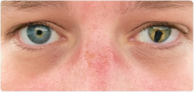
Colobomas are congenital defects of ocular tissues that result in a gap or hole in some structure within the eye. They may affect any part of the eye from the cornea to the optic disc.

Coloboma is an eye abnormality that occurs before birth. Colobomas are missing pieces of tissue in structures that form the eye.
How are uveal colobomas treated?
Uveal colobomas are those which affect the iris or choroidal layer. These are covered by retinal pigment epithelium.
Colobomas of the iris normally produce a keyhole iris or an oval iris. Patients may or may not have lowered visual acuity, depending on whether the fovea is affected or not.
The risk of retinal detachment is present in a proportion of patients, often following neovascularization of the choroidal layer. This means that patients with a uveal coloboma must be followed up each year by an ophthalmologist. There are no treatments that can change the coloboma. However, an ophthalmologist can help the patient address visual problems and provide regular screening for eye health.
How are optic nerve colobomas treated?
Not all optic nerve colobomas impair visual acuity, but the spectrum of severity is wide. Some patients may have normal vision while others are unable to see. However, some patients have normal visual acuity initially, but later develop neovascular membranes that result in serous retinal detachment. This is especially common with optic disc pits, optic nerve atrophy, or excavated colobomas.
When the coloboma affects the optic nerve head, there is much more damage than with optic nerve pits. In this case the patient has a pupillary defect, but also lack of mature retinal and retinal pigment epithelium. The lamina cribrosa is also defective, while the scleral canal is larger than usual. These features are dangerous in that they allow communication to occur between the vitreous body, the subretinal and subarachnoid spaces, and the space around the eyeball. This can end in a retinal detachment.
It is difficult to treat retinal detachment in patients who have optic nerve coloboma. In most cases the attempt to close the hole is made with diathermy, laser or cryotherapy, which can be applied only with great care to avoid damage to the optic nerve nearby. However, hole closure is rarely achieved.
Pulling on the vitreous in the coloboma could induce a posterior vitreous detachment, and this could help relieve the traction, promoting retinal reattachment. The chances of recurrence are very high.
How is visual acuity improved?
Early intervention programs are to be strongly advised to preserve maximum vision in all patients.
In these patients, refractive errors need to be corrected using lenses. This must be accompanied by preventing or treating amblyopia (lazy eye). It is necessary to improve the vision of the eye with worse vision when both eyes don’t have the same visual acuity.
Patching or the use of eyedrops that cause blurring of vision in the stronger eye for some time is often recommended.
Genetic testing
Genetic testing is indicated if syndromic coloboma is suspected, and will help detect chromosomal or single-gene defects in some cases. However, it is not useful in isolated coloboma which is typically sporadic.
Despite testing, many patients with multiple anomalies have no known genetic defect, indicating that the cause of many cases is untraceable at present.
Surgery
Surgical management is sometimes needed in the correction of squints, but accommodative hyperopia may result, and this may require corrective spectacles for hyperopia in the good eye. Nystagmus and cataracts also benefit from surgical treatment.
Other measures
Diagnosis of other defects
Any patient with colobomas on both sides should have imaging of the brain and the orbits to rule out the occurrence of midline defects of the brain. In addition, these patients are likely to have photophobia, or eyes which are sensitive to light, and poor vision. They must be treated with sunglasses as well as treatment for anisometric amblyopia.
Another possibility to be evaluated is the presence of syndromic coloboma, associated with defects of the central nervous system, especially if the coloboma occurs with microphthalmos or developmental delay, or the person shows evidence of other congenital anomalies, especially of the kidneys, bones or other organs.
If microphthalmos is present, extensive follow up may be necessary, with the use of prosthetic devices to encourage normal facial growth and especially normal orbit development. Artificial eyes may be useful for cosmetic reasons. The devices used should be changed as growth occurs.
Plastic surgery or colored contact lenses may be offered for large anterior colobomas which affect the appearance of the eyes.
Detection of complications
In all cases the person should be told about the possibility of complications if there is an optic nerve coloboma, namely, visual loss via retinal detachment or choroidal neovascularization. Early detection and treatment of these issues is a priority.
The use of safety glasses during independent activity is advised for any situation not requiring high visual acuity. Goggles must be donned for sports.
Genetic counseling should be offered to patients and family members.
Further Reading
Last Updated: Mar 20, 2019