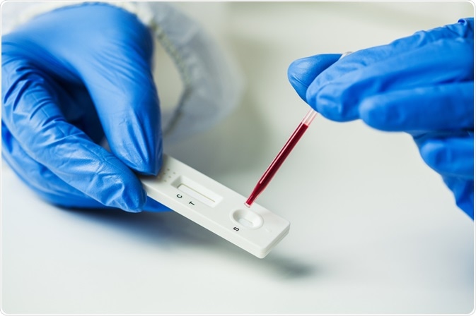
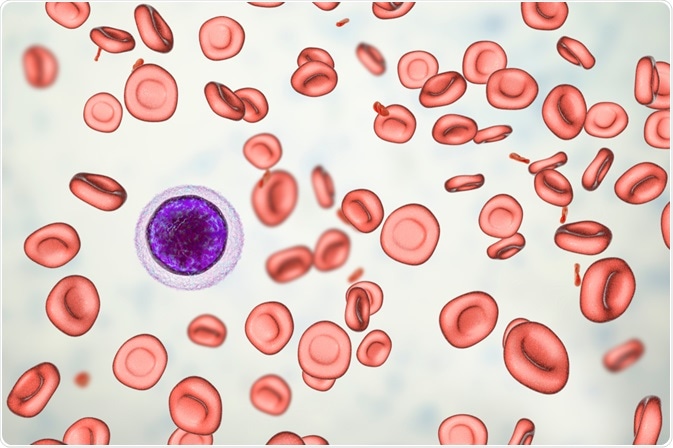
It has been reported that 30% of the world’s population suffers from an iron imbalance. Many diseases such as anemia, hemochromatosis, Parkinson’s disease, cancer, and Alzheimer’s disease can occur due to chronic iron imbalance, i.e., either the concentration of iron in the blood is too high or too low.
 Image Credit: Cryptographer / Shutterstock.com
Early detection and frequent monitoring of iron levels can save patients from permanent disorders, such as impaired developments in children. This article discusses the development of ultrasensitive nanosensors for the detection of iron imbalances.
Common diseases that occur due to iron imbalances
Excess iron concentration can accelerate the aging process. This is because it catalyzes the production of radical oxygen species, which is responsible for harmful oxidative stress leading to cell damage, DNA mutagenesis, and lipid peroxidation.
On sensing a potential threat in our body, an adequate amount of iron is made available to red blood cells. The remaining iron gets converted to ferritin and thus iron is not available to harmful invaders which could have potentially contributed to their growth.
This phenomenon results in a temporary decline in the hemoglobin level. It must be noted that this condition occurs only at the onset of an infection or inflammation and gets rectified once the patient is cured.
The main function of iron in the body is the transportation of oxygen in the blood. Iron deficiency is a condition when the blood contains a low concentration of healthy red blood cells. If this condition is not detected and treated at an early stage, iron deficiency anemia (IDA) may develop.
One of the diseases related to iron overdose is hemochromatosis. Hemochromatosis is regarded as a common hereditary disease. It is a medical condition where patients absorb a high concentration of iron from their diet.
Over a period of time, patients accumulate a high concentration of iron that causes damage to many organs including the liver. Several other diseases that can occur due to iron overdose are osteoporosis and osteopenia, atherosclerosis, Alzheimer's and other neurodegenerative diseases, sarcopenia, metabolic syndrome, etc.
Some of the Nanosensors Developed to Detect Iron Imbalances
Carbon-based Fluorescent Bio-Nanoprobe Technology
Pooria Lesani, a Ph.D. student in the School of Biomedical Engineering at the University of Sydney, has recently developed a novel carbon-based fluorescent bio-nanoprobe technology for rapid detection of free iron ions.
This multipurpose nanoscale bio-probe has the ability to accurately monitor iron disorders in cells, tissues, and body fluids at a very minute concentration, i.e., 1/1000th of a millimolar. The carbon-based fluorescent bio-nanoprobe technology is a non-invasive procedure, that involves subcutaneous or intravenous injections.
Lesani said that this sensor could be used for deep-tissue imaging, where the small probe could visualize the structures of complex biological tissues and synthetic scaffolds.
Most of the available testing approaches are complex and time-consuming. This technology can act as an efficient alternative as it is cost-effective, highly sensitive, non-invasive, and helps in the early detection of serious iron disorder diseases at a very low cellular level concentration.
Lesani and his colleagues tested their technology on the skin of a pig, where they found that the performance of their nanoprobe was much greater than the current techniques available for deep tissue imaging.
These nanoprobes penetrate the biological tissues rapidly to the depths of 280 micrometers and can remain detectable even at depths of up to 3,000 micrometers in synthetic tissue.
At present, the team is focusing on test the activity of the nanoprobes in larger animal models and investigate other modes of application of the nanoprobe for the analysis of structures of complex biological tissue.
Further, the team of researchers at the University of Sydney is planning to integrate the nanoprobe into a “lab-on-a-chip” sensing system. This system would be easy to operate and would require a small quantity of blood samples for the accurate assessment of potential ferric ion disorders in a patient.
Thereby, the development of this portable diagnostic blood testing tool could allow clinicians to remotely monitor their patients’ health.
Graphene-based Biosensor
Researchers have built a graphene-based field-effect transistor (GFET) using graphene monolayer as the conducting channel for early detection of iron deficiency. This channel is activated using anti-ferritin antibodies to selectively trap the ferritin protein antigen. This technology is rapid, straightforward, and inexpensive.
Graphene, besides having many unique properties, is also easy to synthesize. It is inexpensive and extensively commercially available. Iron is detected most accurately by assessing the combination of indicators such as ferritin.
Ferritin is known as an acute-phase reactant protein, whose concentration rises in the presence of infection or inflammation. Thus, ferritin is regarded as a major iron-storage molecule and is widely used as an iron status indicator.
 Image Credit: Kateryna Kon / Shutterstock.com
Electrochemical Nanosensor for the Detection of Hemozoin
Malaria is a complex, infectious, and hematologic disease caused by Plasmodium sp. which is a protozoan parasite. The life cycle of this parasite is partly in humans and partly in Anopheles mosquitoes. Their growth and enzymatic metabolic pathways are dependent on iron.
When humans are infected with this parasite, they degrade hemoglobin. This results in the release of free heme or iron protoporphyrin IX (FePPIX) along with oxygen. Because free heme is toxic to the parasite, it converts the reactive heme species into hemozoin which is nontoxic to the parasite. Thus, hemozoin is known as malaria pigment. It is a visible marker similar to a distinctive hematin pigment, called beta-hematin which also serves as a biomarker for the diagnosis of malaria.
Scientists have developed electrochemical nanosensors using metal oxide nanoparticles which are supported on a gold electrode. This nanosensor has the ability to detect and quantify beta-hematin.
These above-mentioned nanosensors promise to be efficient tools for the detection of analytes of interest in medical diagnostics due to their high sensitivity and selectivity, rapid response time, and low cost.
References
University of Sydney. (2020). Ultra-precision nano-sensor could detect iron disorders. ScienceDaily. www.sciencedaily.com/releases/2020/04/200429105850.htm
Lesani, P. et al. (2020). Two-Photon Dual-Emissive Carbon Dot-Based Probe: Deep-Tissue Imaging and Ultrasensitive Sensing of Intracellular Ferric Ions. ACS Applied Materials & Interfaces, 12 (16): 18395 DOI: 10.1021/acsami.0c05217
Oshin, O. et al. (2020). Graphene-Based Biosensor for Early Detection of Iron Deficiency. Sensors. 20 (13), 3688; https://doi.org/10.3390/s20133688
Obisesan O.R., et al. (2019). Development of Electrochemical Nanosensor for the Detection of Malaria Parasite in Clinical Samples. Frontiers in Chemistry. 7, 89. DOI=10.3389/fchem.2019.00089
Further Reading
Last Updated: Jan 11, 2021