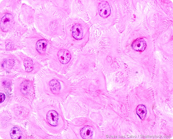
Cadherins are a group of proteins that help cells stick together. They are the main components of certain types of junctions between cells. These connections help define how a cell will be integrated into a structure, like a layer of skin or an organ. Within the junctions, cadherin molecules on the cell surfaces hook onto each other, like a tiny piece of velcro.
Cell-to-cell adhesion mediated by cadherins plays an important role in tissue differentiation, holding the tissues together as they form during embryonic development. Loss of function of cadherins has also been linked to cancer, and some researchers are investigating cadherins as drug targets.
Cadherin structure
Cadherins are named for calcium dependent adhesion. The external domain of a cadherin molecule - the part that is on the outside of a cell - is made up of many repeats of the same protein chain. Each repeat has a space for binding calcium. Calcium makes the chain rigid, helping it to connect with a chain from another cell.
In addition to an extracellular (outside the cell) component, cadherins also have a piece that penetrates through the membrane and a piece that is inside the cell. The interior portions of the cadherins form a complex within the cell.
Types of cadherins
There are about one hundred types of cadherins in vertebrates, and they fall into four groups.
- Classical cadherins include E-cadherin, N-cadherin, P-cadherin and N-cadherin 2. They all have a similar structure, with five extracellular cadherin repeats, a transmembrane domain, and an intracellular domain. Adhesion by classic cadherins is involved in some significant cellular signaling pathways, including Wnt, Hedgehog, Ras, and RhoGTPase signaling. This means cadherins have a critical role in many biological processes.
- Desmoglein and desmocollin are desmosomal cadherins. They are important in forming a type of cellular junction called a desmosome.
- Protocadherins are a large group of cadherin molecules present in a wide range of species that are thought to be related to an ancestral cadherin. Their extracellular domains have more than five repeated cadherin motifs, distinguishing them from classical cadherins. The intracellular domains of the protocadherins are also different from their classical cousins. They are highly variable, with a variety of functions in the nervous system, including neuronal differentiation and the formation of synapses.
- Unconventional cadherins are a large group of cadherins that are not otherwise categorized into the previous three groups. They include VE-cadherin, R-cadherin, and many others.

Cellular junctions rely on cadherins
Two types of cellular junctions rely on cadherins. Aherens junctions have a strip of cadherin molecules that connect the membranes of epithelial cells. The epithelium is a thin tissue that forms layers on hollow structures of the body, like the intestine. Desmosomes also rely on cadherins. A desmosome is a structure inside the membrane of a cell. It uses the cadherins, desmoglein and desmocollin to penetrate through the membrane and form an interlocking network that binds cells together.
In vertebrates, cadherins are responsible for holding most epithelial sheets together. This includes the linings of the small intestine or kidney tubules. Monoclonal antibodies have been used to remove cadherins in cultured epithelial cells, and those cells fall apart. As well, removing calcium from the media will prevent the cell junctions from forming properly.
Cadherins' role in cancer
E-cadherin, a member of the cadherin family, has a very important role in organizing the epithelium. Most cancers arise from epithelial tissue, and in those cancers, cell adhesion mediated by E-cadherin is lost at the same time that a tumor progresses toward malignancy. Some studies have focused on restoring E-cadherin's function in the epithelium as a mechanism for cancer therapy.
Cell organization
Cadherins are also important in other aspects of cell organization, like the formation of boundaries, coordinated cell movements, and maintenance of structural and functional cell and tissue polarity. In addition to E-cadherin's role in the formation of epithelium, N-cadherin is important in neural tissue and muscle, R-cadherin is used in brain and bone tissue, P-cadherin is present in skin, and VE-cadherin is also significant in the epithelium.
Cadherin
Sources
- Nature Reviews, Cell adhesion and signalling by cadherins and Ig-CAMs in cancer, http://www.nature.com/nrc/journal/v4/n2/full/nrc1276.html
- PNAS, Distinct calcium-independent and calcium-dependent adhesion systems of chicken embryo cells, http://www.ncbi.nlm.nih.gov/pmc/articles/PMC319058/?page=1
- Molecular Cell Biology. 4th edition. https://www.ncbi.nlm.nih.gov/
- Expert Opinion on Investigational Drugs, N-cadherin as a therapeutic target in cancer, www.tandfonline.com/.../13543784.16.4.451?journalCode=ieid20
- Tumour Biol., The clinicopathological significance and potential drug target of E-cadherin in NSCLC, http://www.ncbi.nlm.nih.gov/pubmed/25758052
- Cell Mol Life Sci, The cell-cell adhesion molecule E-cadherin, http://www.ncbi.nlm.nih.gov/pubmed/18726070
- Genes and Development, Cadherins in development: cell adhesion, sorting, and tissue morphogenesis, http://genesdev.cshlp.org/content/20/23/3199.long
- Neural Stem Cells - New Perspectives, www.intechopen.com/.../cell-adhesion-molecules-in-neural-stem-cell-and-stem-cell-based-therapy-for-neural-disorders
Further Reading
Last Updated: Jan 2, 2023