BEEC
Cause and symptoms
Epidemiology
Diagnosis and treatment
References
Further reading
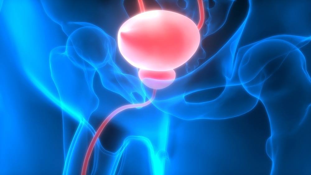
Bladder exstrophy-epispadias-cloacal exstrophy complex is the most severe form of the BEEC (Bladder exstrophy-epispadias complex), constituting 10% of all the cases. In this condition, an abdominal wall defect causes bladder and large intestine extrusion, leading to anal, genital, and colon abnormalities. BEEC is a rare, congenital, multisystemic condition and involves anomalies in the musculoskeletal, urinary, and genital systems. The majority of treatment of BEEC is surgical, and it is usually done in phases. Several long-term consequences of this difficult pediatric urological condition necessitate a comprehensive approach to therapy.

Image Credit: Magic mine/Shutterstock.com
BEEC
BEEC is a term that describes a range of anterior midline defects, from glandular epispadias to multisystem defects like cloacal exstrophy. The word exstrophy comes from the Greek word "ekstriphein," which means "turn inside out." Within the BEEC spectrum, there are three unique conditions. These are - classic bladder exstrophy, epispadias, and cloacal exstrophy.
Classic bladder exstrophy is the most common presentation form of BEEC, constituting 60% of all cases. It is defined by an open, inside-out bladder and an exposed dorsal urethra on the lower abdomen wall's surface. The umbilicus is positioned lower, and pubic bones on both sides of the bladder template can be felt. External genitalia involvement is present in all patients, and most have a palpable inguinal hernia.
BEEC's mildest manifestation is epispadias. It is characterized by non-closure of the urethral plate and an atypical dorsal urethral position, accounting for 30% of all BEEC cases. Males have an ectopic meatus or a mucosal strip on the penile dorsum, whereas females have a urethral cleft.
Cause and symptoms
The specific process that causes bladder exstrophy in the embryo is unknown. The urogenital membrane is thought to break down prematurely because the lower abdominal wall fails to develop. The outcomes include an open bladder plate, a low-placed umbilicus, and a diastasis of the pubic bones. Above the genital tubercle, the cloacal membrane ruptures, resulting in a penis with an open dorsal surface that is continuous with the bladder plate. The type of defect is determined by when the rupture occurs, resulting in the different forms of BEEC.
A mix of genetic and environmental factors has also been discovered to play a role in the disease's etiology. Male sex, race, parental age, and pre-conceptional mother exposure to smoking, certain drugs, and alcohol have all been linked to an elevated incidence of BEEC in studies.
Cloacal exstrophy typically affects the pelvis. Eversion of the innominate bones and widening of the pubic symphysis are frequent symptoms. The pelvic abnormalities in cloacal exstrophy are more severe and asymmetric, and they can result in hip dislocation, necessitating an ultrasonographic examination of the hip joints. In bladder exstrophy and as part of the OEIS syndrome (omphalocele, cloacal exstrophy, imperforated anus, and spinal defects), spinal anomalies such as neural tube malformations can arise.
Several other skeletal and limb malformations are frequent in affected children, especially with cloacal exstrophy, and should be considered during initial monitoring and long-term management planning.
Cloacal exstrophy is almost often linked to gastrointestinal abnormalities. Omphaloceles can be identified in cloacal exstrophy in 88-100 % of patients. In up to 46% of instances, gastrointestinal malrotation and short bowel are present, resulting in absorptive problems in 25% of children. Patients with cloacal exstrophy may also have various urologic problems such as ureteropelvic junction blockage, horseshoe kidney, ureteral ectopy, megaureter, and ureterocele in addition to the bladder defect.
Epidemiology
The incidence of BEEC has been recorded in a variety of ways, including subtypes, ethnicity, and sex ratio. Cloacal exstrophy is the rarest form of the spectrum, with a prevalence of 0.5 to 1 per 200,000 live births. The prevalence of cloacal exstrophy-related pregnancies could be as high as 1 in 10,000 to 1 in 50,000 due to greater rates of stillbirth and pregnancy termination.
Diagnosis and treatment
Clinical observation after delivery is used to diagnose bladder exstrophy in children. Bladder exstrophy, on the other hand, may usually be detected between the 15th and 32nd weeks of pregnancy with high-resolution real-time ultrasonography, even during regular obstetric treatment. The absence of a fluid-filled bladder, a low-set umbilicus, tiny genitalia, pubic rami expanding, and rising lower abdominal mass as the pregnancy progress have all been found to be reliable criteria for prenatal diagnosis of bladder exstrophy.
Only ambiguous instances are subjected to fetal magnetic resonance imaging (MRI) and color Doppler ultrasonography. Although prenatal intervention is not required, early detection allows for delivery in a pediatric center equipped to handle this difficult deformity and thorough family counseling.
Bladder Exstrophy: A Multi-Institution Approach
In bladder exstrophy, treatment aims to close the bladder and abdominal defect while maintaining renal and sexual function. A pelvic osteotomy is always recommended in cases of failed exstrophy repair or cloacal exstrophy. Cloacal exstrophy is more difficult to treat surgically, and it usually necessitates neurosurgery, gastrointestinal, and urological treatments.
Repair of spinal dysraphism and myelocystocele, if present, is included. It also includes repair of the omphalocele, external genitalia, and anorectal malformations, along with the closure of the bladder, urethra, and bony pelvis. However, urinary continence in these children is usually achieved with bladder augmentation and intermittent catheterization.
References
Further Reading
Last Updated: Aug 15, 2022