Causes and pathophysiology
Symptoms and diagnosis
Treatment and management
LAM in perspective: statistics and global impact
Future directions and research
References
Further reading
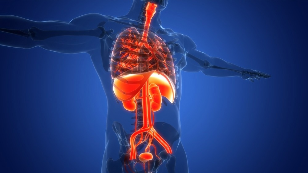
A rare neoplastic condition, lymphangioleiomyomatosis or LAM, is associated with abnormalities in the lungs and kidneys. It is a multisystemic disease linked with TSC1 (tuberous sclerosis complex) and TSC2 mutations commonly observed in young women. LAM patients exhibit a range of clinical symptoms, including cystic lung lesions, dyspnea, and recurrent pneumothorax.

Image Credit: Magic mine/Shutterstock.com
The first case of LAM was recorded in 1918 after a young female died due to complications of tuberous sclerosis and bilateral pneumothorax. Later, in 1937, the first sporadic case of LAM was observed in a female who died due to respiratory complications. Following this, the clinical characteristics of LAM were discussed in 1975, and cases of LAM complications were reported in the 1990s from different parts of the world.
Causes and pathophysiology
Two different forms of lymphangioleiomyomatosis- sporadic and inherited- have been described in the literature. Sporadic LAM is associated with mutations in the TSC2 genes, while the inherited form may occur due to mutations in the TSC1 and TSC2 genes.
These tuberous sclerosis complex genes are responsible for the production of proteins (hamartin and tuberin) that inhibit the target of the mTOR (mechanistic target of rapamycin) signaling pathway. This pathway plays a crucial role in regulating the size and proliferation of cells.
It is a progressive chronic condition, categorized as a low-grade metastasizing lung neoplasm by the World Health Organization (WHO). The abnormal hyperplastic smooth muscle cells that proliferate around and along lymphatic channels, blocking lymphatics, veins, and airways, are the pathogenic hallmarks of LAM.
Neoplastic smooth muscle-like cells, or LAM cells, proliferate in minute clusters at the margins of cysts as well as along blood arteries, lymphatics, and bronchioles in lung lesions. Venous occlusion and bleeding, vascular wall thickening, disruption of the lymphatic arteries, and obstruction of the airways are brought on by LAM cell infiltrates.
Angiomyolipomas are tumors made up of abnormally differentiated cells with three dysmorphic components that resemble vascular, smooth muscle, and adipocyte cells. They manifest as distinct individual tumors that vary in size from microscopic to over 20 centimeters in diameter within the normal kidney or liver parenchyma.
Chyle-filled, encapsulated lymphatic masses, known as lymphphangioleiomyomas, are typically found in the posterior mediastinum, pelvis, and retroperitoneum. LAM cells grouped in fascicular, trabecular, and papillary shapes with slit-like vascular channels make up these tumors.
Symptoms and diagnosis
LAM patients can present with a range of clinical symptoms. Pulmonary symptoms, including breathlessness, pneumothorax, progressive dyspnoea, cough, chest pain, and chylous pleural effusions dominate the clinical course. Recurrent pneumothorax and dyspnoea are highly common, while chylous effusion and hemoptysis are less common. Around half of the patients exhibit angiomyolipoma, a benign tumor of the abdomen.
A tissue biopsy of the lung or affected lymphatics is the gold standard for diagnosing LAM. In LAM, immunohistochemical staining for the melanoma-related antigen HMB45 and the smooth muscle marker actin will be positive. Imaging studies, including CT scans, form the basis of LAM diagnosis.
A thorough review of medical history and examination of pulmonary functions (expiratory flow rates and lung diffusion) capacity can confirm the diagnosis and aid in monitoring the progress of the condition. However, since the early signs may suggest the possibility of other conditions like asthma and emphysema, the diagnosis of LAM can be challenging.
Treatment and management
Research exploring subsequent clinical trials of mTOR inhibitors, such as sirolimus and everolimus, which have stabilized lung function, regressed AMLs, and reduced chylous effusions, has contributed to an improved quality of life in LAM patients. On the other hand, if treatment is discontinued, lung function deteriorates, and AMLs reappear. Furthermore, lung transplantation continues to be the best course of treatment for patients with advanced conditions.
Since exogenous estrogen can worsen LAM, which typically affects pre-menopausal females, different antioestrogen techniques have been employed in the therapy of LAM. In addition to looking for indicators that direct treatment, the hormonal therapy approach should be more carefully defined based on each patient's unique clinical features, menopausal state, pregnancy, estrogen exposure, and exacerbation.
LAM in perspective: statistics and global impact
LAM is frequent in young females who are in their reproductive or child-bearing age. According to several studies, the frequency of LAM in women is roughly 5/1,000,000. Patients with LAM have a median survival of more than 20 years after diagnosis.
The inherited form can affect up to 80% of women with TSC, and the sporadic form affects 3.3-5.7/million women. In addition, 13–38% of men with TSC may develop LAM. A more recent study revealed that the prevalence of LAM in women rises with age and may reach 80%, even though the frequency in women with TSC had previously been thought to be around 26%.
Male sporadic LAM is very uncommon; 10% of men with TSC have cystic lung abnormalities that are consistent with LAM. Male TSC patients (13%) had a lower prevalence of cystic abnormalities in their lungs than female TSC patients (42%).
Future directions and research
Over the last two decades, research on LAM has advanced significantly. This includes the identification of the genetic basis of the disease, the role of the mTOR pathway, and preclinical studies highlighting the potential of mTOR inhibitors. The origin of LAM tumor cells is a crucial subject that needs more research to determine whether the uterus is the source of these cells. Although sirolimus has been utilized successfully, further therapeutic approaches are desperately needed.
Tests are being conducted on novel therapies, such as hydroxychloroquine and RhoA GTPases, such as simvastatin. Other pathways in the pathophysiology of LAM, such as those involving chemokines, metalloproteinases, and lymphangiogenic growth factors, may have prospective therapeutic approaches.
References
- Gibbons, E., Minor, B. M. N., & Hammes, S. R. (2023). Lymphangioleiomyomatosis: where endocrinology, immunology and tumor biology meet. Endocrine-related cancer, 30(9), e230102. https://doi.org/10.1530/ERC-23-0102
- Xu, K. F., Xu, W., Liu, S., Yu, J., Tian, X., Yang, Y., Wang, S. T., Zhang, W., Feng, R., & Zhang, T. (2020). Lymphangioleiomyomatosis. Seminars in respiratory and critical care medicine, 41(2), 256–268. https://doi.org/10.1055/s-0040-1702195
- O'Mahony, A. M., Lynn, E., Murphy, D. J., Fabre, A., & McCarthy, C. (2020). Lymphangioleiomyomatosis: a clinical review. Breathe (Sheffield, England), 16(2), 200007. https://doi.org/10.1183/20734735.0007-2020
- Taveira-DaSilva, A. M., & Moss, J. (2016). EPIDEMIOLOGY, PATHOGENESIS and DIAGNOSIS of LYMPHANGIOLEIOMYOMATOSIS. Expert opinion on orphan drugs, 4(4), 369–378. https://doi.org/10.1517/21678707.2016.1148597
- Moir L. M. (2016). Lymphangioleiomyomatosis: Current understanding and potential treatments. Pharmacology & therapeutics, 158, 114–124. https://doi.org/10.1016/j.pharmthera.2015.12.008
- Taveira-DaSilva, A. M., & Moss, J. (2016). EPIDEMIOLOGY, PATHOGENESIS and DIAGNOSIS of LYMPHANGIOLEIOMYOMATOSIS. Expert opinion on orphan drugs, 4(4), 369–378. https://doi.org/10.1517/21678707.2016.1148597
- Taveira-DaSilva, A. M., & Moss, J. (2015). Clinical features, epidemiology, and therapy of lymphangioleiomyomatosis. Clinical epidemiology, 7, 249–257. https://doi.org/10.2147/CLEP.S50780
Further Reading
Last Updated: Dec 4, 2023