Introduction
History
Cause and symptoms
GJA1 gene
Epidemiology
Case reports
Diagnosis and treatment
References
Further reading
Oculodentodigital dysplasia (ODDD) is a rare congenital malformation associated with eye, face, and limb abnormalities. ODDD follows the autosomal dominant pattern of inheritance and develops due to mutations in the GJA1 gene.
This multisystemic genetic disorder involves a spectrum of clinical symptoms. Some common features observed in patients are microphthalmia, congenital cataract, syndactyly, and early tooth loss.
Some characteristics of oculodentodigital dysplasia are visible from birth, whereas others emerge as the patient grows older. Clinical examination and genetic tests can be used to detect oculodentodigital dysplasia.
 Limb abnormalities and complete syndactyly of the fingers are symptoms of oculodentodigital dysplasia. Image Credit: JorgeMRodrigues/Shutterstock
History
Lohmann is considered to have been the first to describe ODDD in 1920. In 1964, a brother and sister with bilateral microphthalmia, an abnormally small nose, dental abnormalities, syndactyly of the fourth and fifth digits, and missing toe phalanges were described by Gillespie. Meyer-Schwickerath et al., who named the condition oculodentodigital dysplasia, had described comparable findings in two unrelated cases in 1957, according to Gillespie.
Rajic and De Veber, in 1966, described a three-generation family with several afflicted members but no male-to-male transmission. In novel mutations for this disorder, Jones et al. (1975) discovered evidence of a paternal age effect.
Cause and symptoms
ODDD is caused by a mutation in the GJA1 gene located on chromosome 6q22-q24 and is inherited in an autosomal dominant pattern. However, sometimes ODDD can be inherited in an autosomal recessive pattern.
The eyes, dentition, and digits are involved with the oculodentodigital syndrome, which leads to a distinct facial expression. Microphthalmia and other eye abnormalities that might lead to visual loss are common in patients with this condition. Complete syndactyly of the fourth and fifth fingers (syndactyly type III) is the most frequent digital deformity, but the third finger can also be affected, and accompanying camptodactyly is prevalent.
Tooth abnormalities, such as undersized or missing teeth, thin enamel, many cavities, and early tooth loss, are common in those who are affected.
Some people who are affected have neurological issues such as bladder or bowel control issues, ataxia, hearing loss, spasticity, and dysarthria. Craniofacial characteristics such as a narrow nose, hypoplastic alae with a lengthy nasal bridge, cleft lip and palate, and mandibular overgrowth are also observed. Occasionally, ODDD patients may also exhibit brachydactyly, brittle hair, epicanthus, hearing impairment, and intellectual disability.
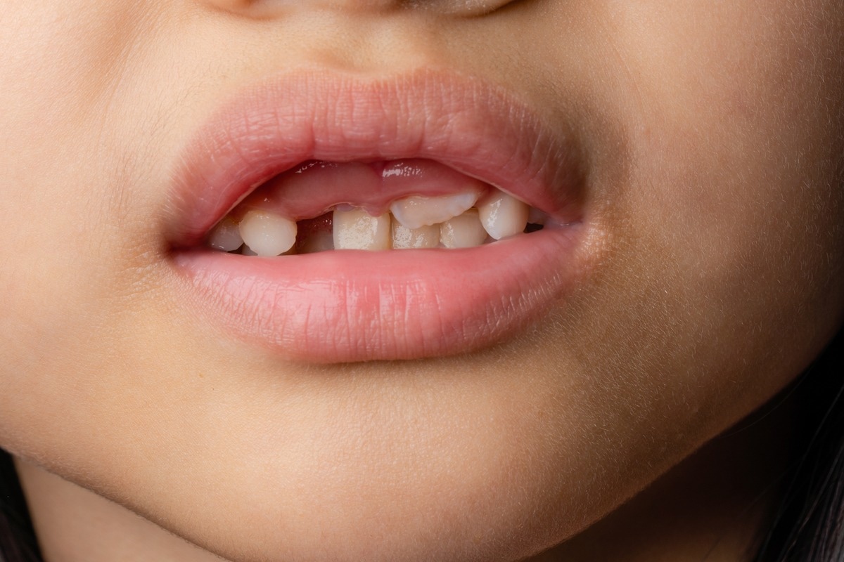
 Early tooth loss is common with ODDD. Image Credit: Michaelnero/Shutterstock
GJA1 gene
The GJA1 gene regulates the production of a protein known as connexin 43. This protein is a component of gap junctions, which are channels that allow direct communication between cells. Connexin 43 proteins produce gap junctions in a variety of tissues throughout the body. Connexin 43 proteins are dysfunctional when the GJA1 gene is mutated. Abnormal protein-formed channels are often permanently blocked.
Connexin 43 proteins are unable to move to the cell surface, where they are required to establish channels between cells, due to mutations. Cell-to-cell communication is disrupted by faulty channels, which may interfere with normal cell development and specialization, processes that dictate the shape and function of many different body parts. The signs and symptoms of oculodentodigital dysplasia are caused by these developmental issues.

 Read Next: What are Genetic Disorders?
Epidemiology
It has only been diagnosed in about 300 patients around the world, with a 1 in 10 million occurrence rate. ODDD affects both males and females in equal numbers.
Case reports
Chai et al., (2019) presented a 23-year-old woman with characteristic ODDD facial symptoms. The hair and brows were thin and fragile, according to a routine physical assessment. When it came to the oral examination, the teeth had a premature appearance due to microdontia. They performed epicanthoplasty, lateral canthotomy, and correcting treatments for both sides of the 3rd and 4th fingers syndactyly at the same time due to the patient's age, comorbidities, and personal facial condition.
The physicians also applied the Rosenberg self-esteem scale (RSES) to assess the patient's self-esteem before surgery, 9 days following surgery (suture removed), and 1 month after surgery. Self-esteem was higher in the postoperative stage, according to the findings. The cosmetic results were also satisfactory to the patient.
Shinya et al. (2021) described a 42-year-old carpenter who had a history of migraine and bilateral syndactyly, which manifested as numbness in the limbs and shaking legs, preventing him from working. A neurological examination indicated spastic paraparesis in all four extremities, as well as abnormal reflexes. Based on his medical history and distinctive facial features such as small eye slits, a slender mouth, and a pinched nose with anteverted nostrils, ODDD was suspected.
A GJA1 gene mutation was discovered during genetic testing, confirming the diagnosis of ODDD. His spastic paraparesis was resistant to oral antispastic medication, but after starting intrathecal baclofen therapy, his symptoms improved significantly, allowing him to return to work.
Diagnosis and treatment
Clinical examination and genetic tests can be used to detect oculodentodigital dysplasia. Hallermann–Streiff syndrome, ectrodactyly ectodermal dysplasia clefting (EEC) syndrome, orofacial digital syndrome Type II, and keratitis ichthyosis deafness syndrome are included in differentiating diagnoses.
Multidisciplinary management is required. A full ocular, neurological, hearing, and oral examination should be performed on a regular basis. Severe limb abnormalities may require plastic or orthopedic surgery.
The importance of early diagnosis of the condition in the prevention and treatment of diverse clinical manifestations cannot be overstated. Systemic examination and genetic counseling aid in the diagnosis of the condition and the prevention of worsening symptoms.
 Genetic testing is often used to confirm a diagnosis of ODDD. Image Credit: Evgeniy Kalinovskiy/Shutterstock
References
- Shinya, A., Takahashi, M., Sato, N., Nishida, Y., Inaba, A., Inaji, M., Yokota, T., & Orimo, S. (2021). Oculo-dento-digital Dysplasia Presenting as Spastic Paraparesis Which Was Successfully Treated by Intrathecal Baclofen Therapy. Internal medicine (Tokyo, Japan), 60(14), 2301–2305. https://doi.org/10.2169/internalmedicine.6145-20
- Chai, N., Lang, Z., Wang, M. et al. (2020). Oculodentodigital dysplasia: plastic treatments and self-esteem estimation. Eur J Plast Surg, 43, 657–660. https://doi.org/10.1007/s00238-019-01594-y
- Attig, A., Trabelsi, M., Hizem, S., Ben Jemaa, L., Maazoul, F., Chaouachi, S., & Mrad, R. (2016). OCULO-DENTO-DIGITAL DYSPLASIA IN A TUNISIAN FAMILY WITH A NOVEL GJA1 MUTATION. Genetic counseling (Geneva, Switzerland), 27(3), 433–439.
- Oculodentodigital dysplasia; ODDD. [Online] OMIM. Available at: https://omim.org/entry/164200
- Oculodentodigital dysplasia. [Online] NIH-GARD. Available at: https://rarediseases.info.nih.gov/diseases/7239/oculodentodigital-dysplasia
- Oculodentodigital dysplasia. [Online] Medline Plus. Available at: https://medlineplus.gov/genetics/condition/oculodentodigital-dysplasia/
Further Reading
Last Updated: Jun 23, 2022