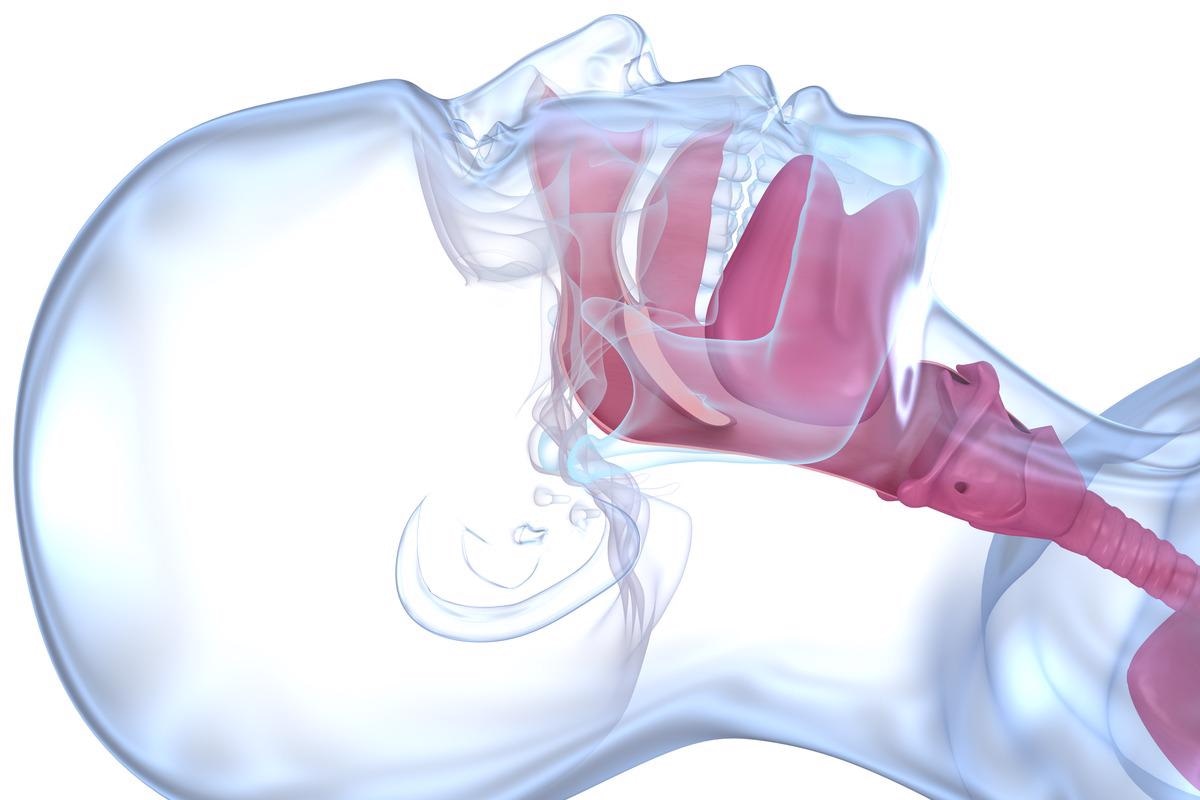
Subglottic stenosis (SGS) is a condition in which the airway below the subglottis and above the trachea narrows. It can be either congenital or acquired.
Subglottic stenosis is a condition that can affect newborns, toddlers, and adults. A developmental failure during pregnancy is thought to be the cause of congenital SGS. It is usually caused by an iatrogenic injury caused by medical treatment or other forms of blunt or penetrating trauma. The severity of the subglottic blockage determines the management strategy in a major part.

Image Credit: Emily frost/Shutterstock
Causes and symptoms
Though trauma or intubation problems can cause subglottic stenosis, the cause of many cases is unknown. Idiopathic subglottic stenosis (iSGS) is the medical term for this condition with no known cause. Although many theories exist that consider genetic, environmental, and immunologic components, there is currently insufficient evidence to provide definitive conclusions.
The subglottis is also thought to be particularly sensitive to instrumentation and pathological processes. The subglottis may be particularly damaged by a shaky blood supply or microcirculation since it connects two embryological growth regions. Other factors that have been linked to stenosis are infection, stomach reflux, radiation therapy, turbulent airflow, and the existence of a full cartilage ring.
Patients suffering from SGS experience various symptoms including dyspnea, recurring croup, stridor, increased mucous production, and change in voice. Infants with SGS may also experience respiratory distress, difficulty in weight gain, and cyanosis.
Grading
The severity of subglottic stenosis is measured using the Cotton-Myer grading system, which compares the appropriate endotracheal tube size to age-matched norms. Grade I signifies up to 50% stenosis and Grade II corresponds to 51-70% stenosis. Grade III corresponds to 71-99% stenosis, whereas Grade IV indicates the absence of lumen.
Types of SGS
Congenital SGS is often linked with other congenital head and neck diseases and syndromes and is due to a developmental failure that occurs during pregnancy. It is the third most frequent congenital defect of the airway. In the absence of prior intubation or airway equipment, it happens when subglottic patency narrows. Congenital subglottic stenosis is a rare birth abnormality that can be linked to various genetic syndromes like trisomy 21, CHARGE syndrome, and 22q11 deletion syndromes. Congenital SGS can be cartilaginous or membranous, and it can range from mild constriction to full laryngeal atresia.
The most frequent acquired laryngeal abnormality is acquired subglottic stenosis, which is caused by prior intubation in 90% of cases. The subglottis' anatomic restrictions make it vulnerable to iatrogenic damage, delayed healing, and stenosis. Preventive measures are therefore necessary to reduce the chance of developing subglottic stenosis.
Idiopathic subglottic stenosis (iSGS) is an uncommon fibrotic condition with unknown etiology and pathobiology that occurs when no cause can be found despite a thorough examination. iSGS is a relatively new entity, with Brandenburg describing the first examples in1972. Only anecdotal accounts and few case studies have been published since then. The cause of this condition is unknown and diagnosing and treating it remains a clinical challenge.
Epidemiology
Subglottic stenosis is a relatively uncommon condition. It affects roughly 1 in every 400,000 people. For iSGS, the majority of patients are Caucasian women. It affects virtually mainly women in their third and fifth decades of life, with an estimated frequency of 1:400,000. The average age is between 30 and 60 years old.

Image Credit: Alex Mit/Shutterstock
Diagnosis and treatment
Clinical history and physical examination play a critical role in the diagnosis of SGS. Investigations about birth injury, intubation, or preterm, as well as other congenital anomalies and physiologic indicators of cardiovascular and respiratory health, should be made in the unintubated nontracheotomy-dependent child. The sound of one's voice is particularly essential because it can reveal the anatomic location of the pathology.
The differential diagnosis includes conditions that induce stridor. Croup, bacterial tracheitis, subglottic cysts, subglottic hemangioma, vocal cord paralysis, and complete tracheal rings are the most prevalent. X-rays are used to further assess the situation. X-rays of the neck may indicate subglottic constriction or masses. The trachea is examined on these films for tracheal constriction, stenosis, or full rings. Endoscopy with microlaryngoscopy and bronchoscopy are used to get a definitive diagnosis.
SGS is currently treated with a variety of endoscopic and open surgeries, and these methods are not always mutually incompatible. Endoscopic techniques are employed as the main treatment and as a follow-up treatment before or after open airway reconstruction. Conservative treatment is employed for mild cases of subglottic stenosis, i.e, for patients with Grade I and II disease. For moderate to severe cases (Cotton-Myer grades III and IV) sometimes surgical intervention and long-term tracheostomy dependency are necessary. Open surgical procedures are reserved for patients with more severe subglottic stenosis or multilayer disease.
Patients with grade II and grade III subglottic stenosis may benefit from laryngotracheal reconstruction. It can be conducted in both single-stage and double-stage manner. Balloon dilatation of the airway with high-pressure noncompliant balloons has advanced significantly in recent years and is now regarded as an important addition to the techniques used to treat SGS, reportedly lowering the need for open airway surgery by up to 80%.
References:
- Aravena, C., Almeida, F. A., Mukhopadhyay, S., Ghosh, S., Lorenz, R. R., Murthy, S. C., & Mehta, A. C. (2020). Idiopathic subglottic stenosis: a review. Journal of thoracic disease, 12(3), 1100–1111. https://doi.org/10.21037/jtd.2019.11.43
- Marston, A. P., & White, D. R. (2018). Subglottic Stenosis. Clinics in perinatology, 45(4), 787–804. https://doi.org/10.1016/j.clp.2018.07.013
- Jefferson, N. D., Cohen, A. P., & Rutter, M. J. (2016). Subglottic stenosis. Seminars in pediatric surgery, 25(3), 138–143. https://doi.org/10.1053/j.sempedsurg.2016.02.006
- Hseu, A. F., Benninger, M. S., Haffey, T. M., & Lorenz, R. (2014). Subglottic stenosis: a ten-year review of treatment outcomes. The Laryngoscope, 124(3), 736–741. https://doi.org/10.1002/lary.24410
- Subglottic Stenosis. [Online] Cleveland clinic. Available at: https://my.clevelandclinic.org/health/diseases/22031-subglottic-stenosis
- Subglottic Stenosis. [Online] Children’s hospital of Philadelphia. Available at: https://www.chop.edu/conditions-diseases/subglottic-stenosis#:~:text=Subglottic%20stenosis%20(SGS)%20is%20a,cartilage%20ring%20in%20the%20airway.
- Subglottic Stenosis. [Online] Nationwide Children’s. Available at: https://www.nationwidechildrens.org/conditions/subglottic-stenosis
Further Reading
Last Updated: May 19, 2022