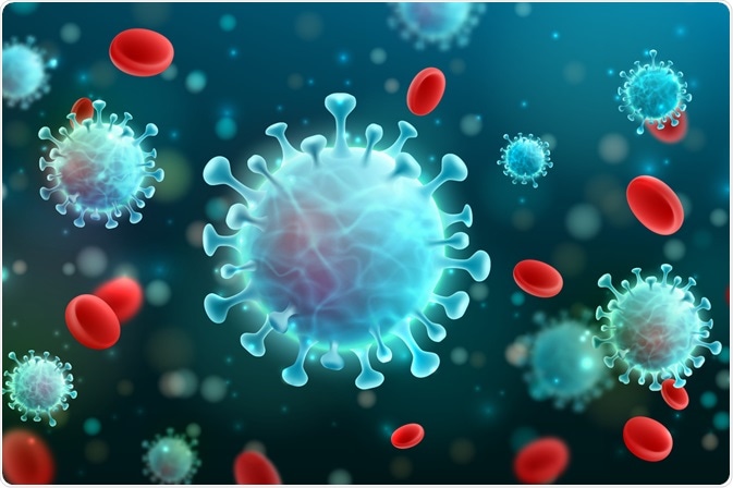
Virotherapy is an emerging branch of medicine that explores the use of viruses to kill cancers.
What is oncolytic virotherapy?
Oncolytic virotherapy can be defined as the elimination of malignant cells by viral propagation inside the tumor. The biological basis of this phenomenon is virus tropism, which means that different viruses have selective preferences for particular cells.
This results in the differences in disease manifestation from virus to virus. Most viruses in nature have a tropism for malignant cells, which makes them suitable for oncolysis. Oncolytic viruses include the oncolytic measles virus, oncolytic vaccinia viruses, oncolytic HSV, reovirus, and much more. In some trials, oncolytic viruses have been combined with chemotherapy and/or radiation.

Image Credit: Fotomay / Shutterstock.com
Normal antiviral responses
The body normally responds to viral infection by slowing or stopping cell metabolism to prevent virus replication, which is typically achieved through the initiation of apoptosis, which is otherwise known as programmed cell death. Tumor cells, however, resist apoptosis and do not suppress protein synthesis following viral infection, thereby making them more susceptible to damage and death.
This is the basis of oncolytic virus therapy. Even attenuated viruses can propagate inside tumor cells, where they otherwise could not survive inside a normal cell. However, the immune responses in most cancer cells are only partially inactivated, so most tumors show only limited sensitivity to viral infection.
To overcome this, virotherapy researchers encode virulence factors that are either complementary to each other or originate from competing virus strains. At the same time, these therapies require some form of protection for normal cells against any potential viral infection.
Mechanisms of oncolysis in virotherapy
The viruses used in oncolysis must be capable of replication and selective infection of tumor cells. Oncolytic viruses use many mechanisms to kill cancer cells, either directly, or through immune mechanisms which destroy the infected tumor cells.
During oncolysis, a virus will utilize cell destructive processes like autophagy, apoptosis, or necrosis after it has fully used up all cell resources to replicate itself. Viruses will also destroy uninfected cells through indirect means, such as disrupting the blood supply to the area, amplifying the existing immune responses against the cancer cell to make the virus more powerful, as well as through specific actions by genetically engineered viruses on the cancer cells.
Update on oncolytic viruses for cancer treatment
The viremic threshold
The two important areas of research into oncolytic viruses concern virus delivery to the tumor and investigating how to spread the virus through the tumor. Effective virus delivery requires the ‘viremic threshold’ dose, which needs to be exceeded in order to be successful.
The safety record of oncolytic virotherapy has thus far been satisfactory. The greatest challenge in virotherapy, however, is making sure that viruses reach the tumor in sufficient doses to extravasate from blood vessels into the tumor. Once inside the tumor, the viruses must successfully replicate in the cancer cells to create infectious centers, which then expand outwards and merge to destroy the tumor, a process that is otherwise known as effective tumor debulking.
Some of the major obstacles encountered in virotherapy include:
- Sequestration of the virus in the liver and spleen, which may be overcome by saturating the mononuclear phagocytic system receptors.
- Neutralization of viruses by serum immune factors.
- The need for viruses to selectively bind to the vascular endothelium of the tumor blood vessels.
- The need for a highly selective increase in the permeability of tumor vascular endothelium to optimize virus extravasation while simultaneously avoiding systemic inflammation.
Reporter genes
The spread of the virus, as well as the number and location of virus-infected cells inside the body, can be tracked by monitoring certain inserted genes. These inserted genes, which are otherwise known as reporter genes, help researchers trace the virus spread repeatedly and in a non-invasive way.
One example of a reporter gene is the gene that encodes the thyroidal sodium iodide symporter (NIS), which concentrates radioactive iodide in the form of radioisotopes such as 125I, 123I, 124I, and 99mTcO4. The NIS gene can be inserted into the virus prior to injection into the patient to allow clinicians to monitor the spread of the virus. This approach has been validated with the use of CT/SPECT imaging to follow the spread of the oncolytic virus with the inserted NIS.
Newer developments and challenges
Radiovirotherapy is another approach under investigation, which uses 131I in the form of NIS expression in a virus genome. This will increase the efficacy of the oncolytic virus therapy by adding the administration of high-energy beta particles into the virus-infected tumor to the virus infection.
The large size of most viruses forms a physical barrier to cell infection, while their ability to evoke an immune response limits their capacity to infect many cells. However, both of these characteristics of viruses can amplify and prime other forms of host immunity and thus stimulate a general antiviral immune response in the host. Cell carriers are being developed to overcome the physical size barrier. The use of immunosuppressive drugs is also being researched to encourage the intratumoral spread of the virus.
Challenges exist within virotherapy research, as the diminishing of host immunity subsequently reduces the immune-stimulating power of oncolytic virus therapy. Moreover, there remains an urgent need to develop tumor models that can accurately reflect what is happening both within the oncolytic virus-infected tumor and the host as a whole.
References
Further Reading
Last Updated: Mar 17, 2021