Functional magnetic resonance imaging (fMRI) is a non-invasive and safe technique to measure and map the activities of brain during normal as well as diseased conditions. It measures the the changes in the brain’s blood flow that happen with brain activity.

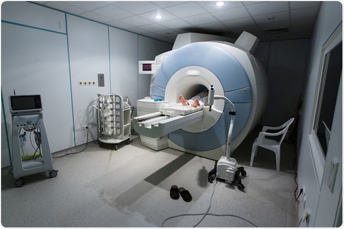
Patient being scanned and diagnosed on a MRI (magnetic resonance imaging) scanner in a hospital. Image Credit: zlikovec / Shutterstock
The fMRI is an extensively used technique in Radiology that produces high resolution images with good contrast between different brain tissues. The basic principle of fMRI is based on the fact that nucleus of a hydrogen atom possesses the property of a small magnet. Functional MRI uses the principle of nuclear magnetic resonance, which is a physical phenomenon wherein certain atomic nuclei present in a strong stationary magnetic field selectively absorb very high-frequency radio waves and produce an electromagnetic signal with a frequency of the magnetic field at the nucleus.
In fMRI, a strong magnetic field is applied to align hydrogen atoms that are otherwise randomly oriented within water nuclei of the tissue of interest. Upon applying a radio frequency magnetic pulse at an appropriate frequency, these nuclei absorb energy and produce a signal (magnetic resonance signal) that can be detected by the radio frequency coils present in the MRI setup.
Principles of fMRI Part 1, Module 1: Introduction to fMRI
Different types of images of a selected brain area can be obtained by changing the sequence of applied and collected radio frequency pulses. The duration between successive pulse sequences applied is called Repetition Time (TR), and the duration between application of the pulse and collection of the signal is called Time to Echo (TE).
In fMRI, two different relaxation times, T1 and T2, are used to characterize a tissue. Longitudinal relaxation time or T1 is the time constant that measures the time taken by excited hydrogen atoms to realign with the external magnetic field. On the other hand, transverse relaxation time or T2 is the time constant that measures the time taken by excited hydrogen atoms to either reach equilibrium or go out of the phase (dephasing). T1-weighted (short TE and TR) and T2-weighted (longer TE and TR) scans are the most common sequences used in fMRI.
How does fMRI brain scanning work? Alan Alda and Dr. Nancy Kanwisher, MIT
Physiological Basis of fMRI
The change in fMRI signal according to altered brain activity is an indirect effect, which depends on the change in cerebral blood flow caused by altered neural activity. An increased neural activity elevates the level of blood oxygenation because of an increased energy demand that subsequently calls for more oxygen. The fact is that oxygen-rich blood and oxygen-poor blood possess different magnetic properties due to the difference in hemoglobin concentration that binds to blood oxygen. When the blood is more oxygenated, the signal is stronger and vice versa. This phenomenon forms the basis of blood oxygenation level-dependent (BOLD) fMRI.
Principles of fMRI Part 1, Module 8: fMRI Signal & BOLD Physiology
The change in blood flow is a very sensitive indicator of altered neural activity. For example, simple tapping of fingers in one hand is capable of increasing blood flow by 60% in the motor area of the brain. Because of this fact, BOLD changes in magnetic resonance signal is sensitive enough to detect subtle changes in neural activity related to simple motor tasks, such as holding a cup of coffee, as well as complex cognitive functions, such as attention, learning, and memory formation.
In basic fMRI, BOLD signal provides only a qualitative overview of changes that are happening due to brain stimulation compared to resting condition. It cannot provide information on actual changes in blood flow before any stimulation is applied.
BOLD contrast fMRI is a useful alternative to overcome this limitation. In this method, a contrast agent is injected in blood to quantitatively detect blood flow at baseline. BOLD contrast is resulted from changes in magnetic field depending on the oxygen state of hemoglobin.
The basic principle is that fully oxygenated hemoglobin is diamagnetic, which is magnetically same as brain tissues. In contrast, deoxygenated hemoglobin is paramagnetic, which leads to generation of local magnetic gradients in and around the blood vessel whose strength depends on the hemoglobin level. The ultimate effect in the magnetic resonance signal depends on the selected field strength, pulse sequence, and time to echo.
This BOLD effect is directly related to the concentration of deoxygenated hemoglobin; highly active brain regions have high concentration of oxygenated hemoglobin and always show higher signals. MRI contrast agents capable of altering oxy/deoxy hemoglobin ratio in brain regions, such as gadolinium, are used to improve the quality of the image by enhancing BOLD contrast. The relaxation time in this case is denoted as T2*, which basically results from the non-uniformity of the main magnetic field resulted from local magnetic gradient. T2*-weighted MRI sequences are used to highlight the magnetic uniformity effects to generate high contrast images through fMRI.
Further Reading
Last Updated: Oct 9, 2018