Introduction
Structural and functional medical imaging
Radiation exposure
Individual techniques
References
Further reading
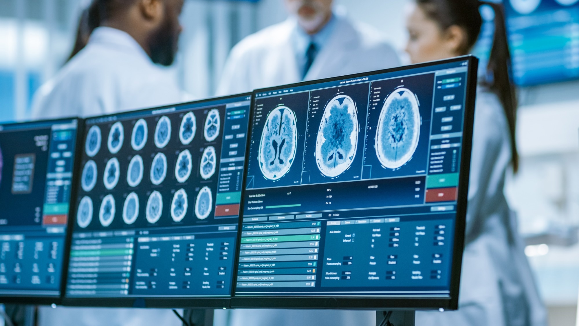
The use of diagnostic imaging in medicine dates back over a century. However, tremendous advances have been made over the last 50 years, in which multiple imaging modalities have offered a previously unimaginable wealth of data on the structure and function of the inward organs of the human body.

Image Credit: Gorodenkoff/Shutterstock
Structural and functional medical imaging
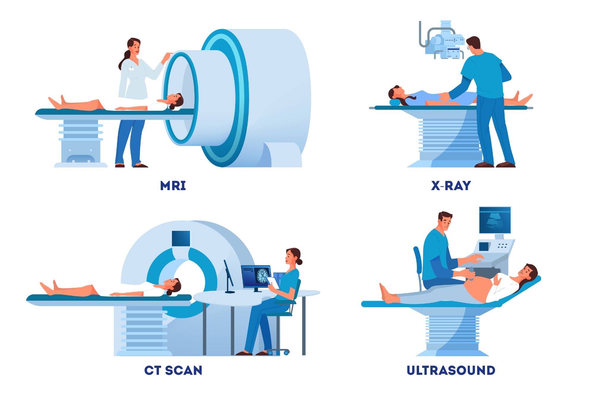
Imaging technologies are directed mainly at elucidating the structure or function of tissues and organs. Structural imaging includes producing images of the anatomy and morphology of the body part generated using computed tomography (CT), magnetic resonance imaging (MRI), and ultrasound (US) scans.
Conversely, the working of a tissue or organ in health and disease is equally vital data for diagnosis and treatment and is called functional imaging. This includes multiple quantitative imaging techniques that look into physiology at the molecular level, using metabolites and other molecules involved in cellular pathways.
Techniques like CT, MRI, and US scanning also allow for functional imaging. However, these are complemented by others, such as positron emission tomography (PET), single-photon emission computed tomography (SPECT), and optical imaging. Whereas the former is better at providing structural and anatomical images, all help to arrive at a good idea of organ function.
However, the spatial resolution of each modality varies, with MRI and US imaging providing a resolution of 50-500 μm and 200 μm, respectively. CT, PET, SPECT, and optical imaging have a resolution of 0.5-1 mm, 1-4 mm, 0.3-3 mm, and 0.1-10 mm, respectively.
The development of contrast agents has helped to provide greater resolution and specificity to the final images, reducing the proportion of noise to signal and allowing both purposes to be fulfilled with the same imaging technique.
Radiation exposure
CT, SPECT, and PET imaging modalities all expose the patient to ionizing radiation, a disadvantage not shared by MRI, US, or optical imaging. On the other hand, MRI is a relatively expensive modality, while the latter two techniques have relatively poor tissue penetration, offering low-quality images beyond the surface.
Together, these are often used to provide more information with lower radiation exposure than one set alone. This is also called multimodal imaging and takes advantage of the complementary aspects of each technique individually. For instance, hybrid PET-MRI scanners are being tested preclinically for their ability to provide dynamic imaging without exposure to ionizing radiation.
The outcome is a more precisely defined knowledge of where a disease process is located, how it affects the organ or tissue, and the patient's health overall. It also helps to decide how best to treat the patient and monitor their health during and after treatment.

 Read Next: Imaging in Nephrology
Individual techniques
Computerized tomography (CT)
CT scanning uses an X-ray beam passed through the body that is partly absorbed or scattered within the imaged organ. The residual X-rays follow multiple linear paths to fall onto a rotating detector, forming a cross-sectional image of the body. While the CT scan uses more radiation than an ordinary X-ray, the volume of the body to be scanned, the number of sequences, and the quality/resolution of the images desired all affect the radiation dose.
Sometimes contrast CT scans are used, with heavy molecules used as dyes to help the soft tissues stand out at more excellent resolution. Iodine-based compounds are used as water-soluble dyes and also to measure renal function. These do not confer color on the organs but change how the imaging beam interacts with them.
Ionic and non-ionic iodine-based dyes are used, with high and low osmolarity. Second-generation compounds based on barium or gadolinium, with lower osmolarity and toxicity than ionic compounds. Unfortunately, these may sometimes trigger allergic reactions in up to 3% with non-ionic dyes but up to 12% with ionic contrast.
CT scanning has come into its own in angiography and other cardiovascular applications. CT angiography (CTA) can show arteries and veins within an organ in a 3D view from any desired angle. Another application is the artery calcium score, which uses CT scans to visualize calcium deposits within the coronary arteries, indicating a higher risk of cardiovascular disease.
Diagnostic Imaging Explained (X-Ray / CT Scan / Ultrasound / MRI)
Magnetic resonance imaging (MRI)
MRI is a technique that uses magnetic fields to align the protons within hydrogen atoms found in water which makes up most of the human body. The MRI scanner emits short radio wave pulses, exciting the protons and breaking up their alignment in the scanned part of the body. As the protons realign, they emit radio waves that are detected to produce high-resolution images of organs and internal structures.
The MRI signal is thus based on the water content and magnetic properties of the body part imaged because the speed of realignment varies from tissue to tissue, producing different signals in each. The millions of radio wave emissions are integrated by a computer to provide a detailed picture of the inside of the organ or the body.
Gadolinium-based non-ionic contrast agents can also be used. Interestingly, the discovery that deoxyhemoglobin is a strongly paramagnetic molecule is used to understand changes in the structure and activity of the brain. The resulting local magnetic field disturbances around and within blood vessels alters the MRI appearances near them.
This has led to the use of functional MRI to detect how tumors, cerebrovascular accidents, brain trauma, and neurodegenerative disorders affect the brain and produce a map of the brain's structure. This is called BOLD (Blood Oxygen Level Dependent) contrast.
MRI does not employ ionizing radiation and is considered among the safest imaging platforms. Researchers have not found harm from using a brief strong magnetic field. However, metal implants, including pacemakers and metal implants, may heat up or fail if exposed to this technique. Pregnancy is a contraindication as well.
Positron emission tomography (PET) and single-photon emission computed tomography (SPECT)
SPECT and PET are both highly sensitive nuclear imaging techniques, where small amounts of radioactive tracers are used to produce images of the body part where they are concentrated. External detectors are responsible for capturing the radiation. These are fed to a computer that generates the image.
The main limitation of these techniques is the poor spatial resolution, as well as, with SPECT, the lack of attenuation that is required to build the image accurately, and the few radiopharmaceuticals available currently. The radiotracers are excreted within 72 hours.
PET is useful in studying metabolically active tissue, its structure, function, and biochemical characteristics. It uses positron emitters, while SPECT uses γ photon emitters, which have slightly longer half-lives. The positrons come from the breakdown of the radionuclide, like fluorodeoxyglucose (FDG), created by attaching a glucose atom to a radioactive label.
The positrons collide with nearby electrons to create gamma rays or annihilation protons. These are detected by the gamma-ray scanners, and the signals are passed to the computer to create an image map displaying the tissue structure and how metabolically active it is.
PET uses a larger range of compounds, expanding its ability to visualize cellular metabolic and functional pathways. The final image is also more accurate with PET because of the corrections applied to the image during reconstruction.
However, all PET tracer molecules have the same decay energy, preventing the distinguishing of different tracers simultaneously. In addition, increased availability of radionuclides and the introduction of gamma camera systems have extended the reach of PET.
Proton magnetic resonance spectroscopy (1H-MRS) is another technique based on the relationship between proton resonance frequency and the chemical environment, lipids, water, amino acids, and the like. This allows evaluation of the brain and metabolic processes during health and disease.

Image Credit: Inspiring/Shutterstock
Ultrasound (US)
US imaging uses sound waves of 1-12 MHz frequency to reflect off internal body structures, thus producing accurate images of such body parts. The underlying principle is the principle of reflection depending on the distance of the reflective surface from the origin of the sound wave. Echocardiography is invaluable in assessing heart size and function, as well as the function of the heart valves.
In contrast, the US has been developed, where gas-filled microbubbles are used intravascularly. They have been proven safe while increasing spatial resolution at a relatively low cost. Doppler US scanning has also proven useful in measuring blood flow through blood vessels.
Optical imaging
Optical imaging uses digital cameras to detect molecular emissions from electromagnetic waves. Without ionizing radiation, it is suitable for repeated examinations, while the addition of contrast can help distinguish between soft tissues. Multiple techniques have been introduced, including diffuse optical tomography (DOT), optical coherence tomography (OCT), and hyper-spectral imaging.
DOT uses low-frequency red and infrared light to detect changes in the concentrations of certain molecules and thus identify changes in cellular function. OCT uses backscattered light detected on the principle of interference and is helpful in examining deeper images. Imaging spectroscopy, meanwhile, combines conventional digital imaging and spectroscopy.
A fascinating advance in this area is molecular imaging, where specific biomolecules that are operative in certain cellular processes underlying one or more disease states are targeted selectively by the imaging modality. Labeled molecules are required, as well as a detector and a ligand to bind to the molecular target, whether this is an antibody, peptide, or small molecule.
Molecular imaging agents are still few, however, and their development is slow. Moreover, the personalized nature of this technique makes its commercialization a less attractive possibility. Nevertheless, the continuing trend towards increased effectiveness and fewer adverse effects in the development of medical diagnostics will probably make multimodal imaging a commonplace entity in various medical conditions.
References:
- Barsanti, C. et al. (2015). Diagnostic and prognostic utility of non-invasive imaging in diabetes management. World Journal of Diabetes. https://doi.org/10.4239%2Fwjd.v6.i6.792. https://www.ncbi.nlm.nih.gov/pmc/articles/PMC4478576/. Accessed on June 23, 2022.
- Hanson, G. (2017). 7 Types of Diagnostic Imaging Tests You May Assist with as a Radiologic Technologist. https://www.rasmussen.edu/degrees/health-sciences/blog/types-of-diagnostic-imaging/. Accessed on June 23, 2022.
- Umar, A. A. et al. (2019). A review of Imaging Techniques in Scientific Research/Clinical Diagnosis. MOJ Anatomy & Physiology. https://doi.org/10.15406/mojap.2019.06.00269. https://medcraveonline.com/MOJAP/a-review-of-imaging-techniques-in-scientific-researchclinical-diagnosis.html. Accessed on June 23, 2022.
- Gilson, W. D. et al. (2022). Noninvasive Cardiovascular Imaging Techniques for Basic Science Research: Application to Cellular Therapeutics. Revista Espanola de Cardiologia. DOI: 10.1016/S1885-5857(09)72656-8. https://www.revespcardiol.org/en-noninvasive-cardiovascular-imaging-techniques-for-articulo-13140623. Accessed on June 23, 2022.
- Baerlocher, M. O. et al. (2010). The Use of Contrast Media. CMAJ. https://doi.org/10.1503%2Fcmaj.090118. https://www.ncbi.nlm.nih.gov/pmc/articles/PMC2855918/. Accessed on June 28, 2022.
- Overview – MRI Scan. https://www.nhs.uk/conditions/mri-scan. Accessed on June 28, 2022.
- BOLD Contrast Mechanism (2021). https://mriquestions.com/bold-contrast.html. Accessed on June 28, 2022.
- Single Photon Emission Computed Tomography (SPECT) (2021). https://www.heart.org/en/health-topics/heart-attack/diagnosing-a-heart-attack/single-photon-emission-computed-tomography-spect. Accessed on June 28, 2022.
- Positron Emission Tomography (PET) (2022). https://www.hopkinsmedicine.org/health/treatment-tests-and-therapies/positron-emission-tomography-pet. Accessed on June 28, 2022.
Further Reading
Last Updated: Sep 29, 2022