What is Optical Coherence Tomography?
Optical coherence tomography (OCT) is a method of optical imaging. It can be used in clinical situations to aid with the diagnosis of diseases in many organs such as the retina, cardiovascular system, gastrointestinal tract, urinary tract, or gynecologic organs.

Optical Coherence Tomography (OCT) image of eye in the patient. Image Credit: Chaiwut Siriphithakwong / Shutterstock
OCT is a high-resolution optical imaging technique that can produce a cross-section image of an object. The image can be produced in real time, allowing this technique to have many applications. There are two methods of OCT: time domain and spectral domain. Within these categories, each one can be performed in different ways. Which one is used depends on the sample being investigated and the advantages and disadvantages of each method.
Applications of Optical Coherence Tomography
Retina
Time domain (TD)-OCT images an object by directly obtaining time-encoded signals. TD-OCT imaging is used to assess the retina to give information regarding retinal thickness and the inner retinal complex (IRC), which is composed of a retinal ganglion cell layer, inner plexiform layer, and inner nuclear layer.
The most common scan patterns of the retina using TD-OCT involves imaging via a 3.4 mm scan around the optic nerve head and six equally spaced radial scans through the macula and optic nerve. This is known as the macular scan pattern. 3D OCT imaging of the retina provides more detailed information than the macular pattern, which can be useful when planning or evaluating the outcome of surgeries. 3D OCT also visualizes the vitreomacular interface in subjects undergoing surgery for epiretinal membrane (a thin sheet of tissue that develops over the macular area, which disturbs vision).
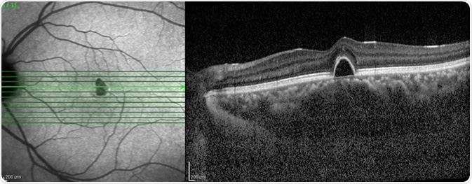
SD-OCT can be used to measure the thickness of the retina and to evaluate the effects of certain diseases on this tissue. The decline in visual acuity seen in diabetic macular edema is caused by retinal thickening. There are many other parameters within the retina that can be measured using OCT such as changes in drusen volume, macular holes, subretinal fluid, and pigment epithelium detachment.
Mastering OCT Interpretation with Dr. Mark Friedberg
Glaucoma
Macular pattern scanning is used to measure the optic nerve head (ONH) and the retinal nerve fiber layer (RNFL) thickness. The RNFL and ONH parameters differ greatly between healthy patients and ones with glaucoma. The screening for glaucoma can be improved by using 3D OCT instead of macular pattern scanning. 3D scanning can reveal the volume of the eye, which then allows for en face images to be obtained using OCT by integrating many individual A-scans. These en face images can be used to assist with ocular evaluations and to correct errors during the scan due to eye motion.
The tests used to analyze the structural differences can only diagnose a disease after a significant difference is observable compared to the healthy phenotype. This presents a problem as some disease conditions fail to show any signs of abnormality, with the structural appearance remaining within the normal parameters. This causes the individual to go undiagnosed. This issue can be reduced by doing longitudinal studies that measure the subtle structural changes that may be due to a disease.
Anterior Segment OCT
Anterior segment (AS)-OCT provides information on the structure of the cornea and anterior chamber of the eye without contacting the eye. This method cannot be used to measure deep tissues within the eye, but it has a very high resolution (5-10 μm) at this level. This makes it possible to acquire high-resolution images of the sclera and the iris. AS-OCT also provides information on corneal thickness.
Cardiovascular System
OCT can be used to diagnose intravascular diseases. To image arteries using OCT, a catheter is inserted into the artery. Blood is removed from the field using a saline flush, which is followed by a helical scan of ~5-10 cm by the OCT catheter’s optics. This provides 3D data on the microstructure of the coronary wall which can be used as a guide during interventional procedures, and can also be used to diagnose atherosclerotic lesions.
The resolution of OCT is high enough to differentiate particles such as calcium, lipids, and thrombi. This allows different atherosclerotic plaque types to be distinguished, further aiding the diagnosis of atherosclerosis.
The Practicality of Optical Coherence Tomography
Optical coherence tomography is an extremely versatile imaging technique. It can be used to image many different organs and organ systems, providing valuable information that can be used to diagnose certain illnesses. This data can also be used to help with the planning and evaluation of surgeries.
As optical coherence tomography becomes more efficient and new ways to use it are devised, it has the potential to improve the diagnosis and treatment of certain diseases.
Further Reading
Last Updated: Oct 14, 2018