By Gillian D’Souza, MSc
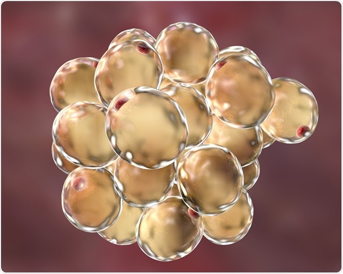
Adipose tissue, commonly known as fatty tissue, is made up of fat cells called adipocytes. Adipo-Clear is a technique used to visualize adipocytes and is used in metabolic experiments to further our understanding of the role of fat in the human body.

Credit: Kateryna Kon/Shutterstock.com
To observe epitopes of interest in adipose tissue, researchers traditionally labelled them on thinly sliced fixed tissues, by one of two methods – immunohistochemistry or immunofluorescence. The high lipid content of these tissues, however, limits the use of these methods, making sectioning difficult and causing light to scatter. With the advent of a new technique called Adipo-Clear, these challenges may soon become a thing of the past.
What is Adipo-Clear?
Adipo-Clear is a new technique used to process and clear adipose tissue for fluorescence microscopy. It allows researchers to visualize 3D images of fat cells and better understand their constituent biology.
The technique was developed by Dr. Paul Cohen and his associates from Rockefeller University in late 2017. Since then, it has shown great potential to overcome limitations posed by traditional histological methods.
How is Adipo-Clear performed?
The Adipo-Clear technique is a modified version of the iDISCO test, developed to immuno-label and 3D visualize key molecular markers in embryonic and adult tissues.
The technique involves de-lipidation and immunolabeling of the fat pads, followed by imaging of the target tissue with the help of light sheet fluorescent microscopy. Adipo-Clear enables complete clearing of lipids in a fat pad, while ensuring that tissue integrity is maintained.
The significance of Adipo-Clear
The extent of knowledge, so far, on adipose tissue biology led researchers to categorize adipose tissue into two forms, namely ‘white’ and ‘brown’. White adipose tissue was found to act as the main site for safe energy storage, while brown adipose tissue dissipated heat energy to protect mammals from hypothermia.
More recently, research has shown that this distinction into white and brown adipose tissue may be too simplistic, given that clusters of UCP1-expressing adipocytes having thermogenic capacity were found in white adipose tissue.
Termed as ‘beige’ adipocytes, these cells develop postnatally in response to certain stimuli or activators. So far, the biology of white and brown fat cells was well understood but limited knowledge existed about beige fat cells and their role in energy use.
Recent developments
Following its development, Adipo-Clear has demonstrated remarkable regional variation in the biogenesis of beige adipocytes within subcutaneous adipose tissue. The technique has highlighted clear differences in the architecture of subcutaneous and visceral fat; the former being organized into discrete lobules and the latter showing signs of amorphous localization. These findings could potentially serve as a guide into better understanding the developmental origins of beige adipocytes.
Adipo-Clear has also made it possible to immunolabel and 3D profile key structures such as thermogenic adipocytes and sympathetic innervation. Given that adipose tissue is a key site for neuronal processes that are critical for thermogenesis, another exciting finding showed a clear relationship between the sympathetic projections in beige adipocytes.
These findings suggest well-defined communication between adipocytes and neurons governed by PRDM-16, a transcription coregulator, and present an opportunity for further research to determine what exactly mediates this relationship.
The introduction of Adipo-Clear has shed more light on beige fat synthesis and its mechanism of action. It is expected that its increased adoption in adipose tissue studies will drive insight into thermogenesis, energy expenditure and metabolism of humans and mammals, and dictate further obesity- and lifestyle disease-related strategies.
Last Updated: Feb 26, 2019