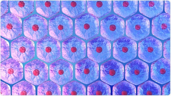
Stem cells contribute to the development of tissues and organs and are responsible for their functioning and maintenance throughout life. They reside in various tissues, and divide to either reproduce themselves or give rise to progeny that undergoes a limited number of divisions and ends up as a functional cell of that tissue.
 Credit: lmstockwork/Shutterstock.com
Exhaustion of the stem cell pool or loss of their ability to produce progeny capable of maturing into functional differentiated cells is currently considered as a main cause of many deceases that accompany aging.
The importance of stem cell labelling
To explore the prospects of healthy tissue rejuvenation or damaged tissue regeneration, it is necessary to uncover the cellular mechanisms supporting the life cycle of stem cells. This means tracking the propagation of stem cells in living tissues, tracing the fate of their progeny, and investigating cell distribution in organs.
Traditional experiments involved tagging the DNA in dividing cells with modified nucleotides. When a cell which has incorporated the tag into its genome divides, all its progenies will be tagged, too. However, so far it has been impossible to use more than two tags simultaneously.
Advances in stem cell labelling
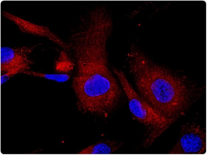
Being able to mark dividing cells with different tags enables cellular processes to be explored in great detail. The problem was that previously, it was impossible to use more than two tags. We recently established a method for labeling three or four DNA tags at once. The use of three or four independent tags allows for marking several cohorts of cells born at distinct timepoints to observe the stem cell progeny passing through the different maturation stages.
Various combinations of pulse and cumulative marking highlights specific subpopulations among dividing stem cells. For example, injecting mice with three tags sequentially revealed the conveyor belt pattern of division and migration of cells in the intestine. Using this technique, we were able to analyze the patterns and parameters of neural stem cell division in the adult brain.
Determination of cycle re-entry, a critical parameter of the cell division kinetics, allowed for the conclusion that neural stem/progenitor cells in the hippocampus, a brain area with continuous neurogenesis, undergo approximately four cell divisions before exiting the cell cycle.
The same approach can be used for parallel analysis of various other dividing cells in an organism. We expect that various combinations of tags and labeling schemes will reveal new complex features of dividing stem cells, such as the distinct steps of their cell cycle entry or exit or the alternation between quiescent and proliferative states, under both normal and pathological conditions.
 Credit: Vshivkova/Shutterstock.com
The future for dividing stem cell labeling
Identifying molecular mechanisms underlying the cell cycle entry or exit or the alternation between quiescent and proliferative states is currently being considered as a critical direction in the field of stem cell research. It is hoped that our technique will contribute to this direction by allowing for identification of stem cells which are undergoing or have recently undergone switch from one state to another.
When combined with the various approaches of genetic tracing, such as Cre-Lox recombination, our method will reveal the proliferative history of definite cell lineages that is critical not only for adult stem cell research, but also for tissue engineering and developmental biology.
About the authors
About Oleg Podgorny
 Dr. Oleg Podgorny is a senior researcher at Laboratory of Brain Stem Cells at Moscow Institute of Physics and Technology, postdoctoral associate at Enikolopov’s Lab at Stony Brook University, and a senior researcher at Laboratory of Regeneration Problems at Koltzov Institute of Developmental Biology RAS.
He received his Ph.D. at Koltzov Institute of Developmental Biology RAS in 2006. He joined Professor Grigori Enikolopov’s group in 2012. His research is focused on elucidating modes of neural stem cell division and understanding mechanisms underlying their maintenance in the adult brain.
About Grigori Enikolopov
 Dr. Grigori Enikolopov is Professor at the Center for Developmental Genetics of Stony Brook University and Adjunct Professor at Cold Spring Harbor Laboratory and at Moscow Institute of Physics and Technology.
He received his Ph.D. at the Institute of Molecular Biology in Moscow. Since 1989 he worked at Cold Spring Harbor Laboratory, before recently joining Stony Brook University. His research focus is on stem cells of the adult brain and signals controlling division, differentiation, and maintenance of stem cells.
Further Reading
Disclaimer: This article has not been subjected to peer review and is presented as the personal views of a qualified expert in the subject in accordance with the general terms and condition of use of the News-Medical.Net website
Last Updated: Apr 12, 2018