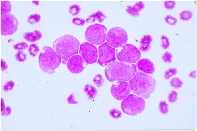
A group from the Shanghai Jiao Tong University School of Medicine has outlined a protocol for the tissue-resident myeloid populations in mouse tissues using flow cytometry.

Image Credit: Medtech THAI STUDIO LAB 249/Shutterstock.com
The protocol builds on existing work by Lu et al. and details the step-by-step procedure for the analysis of myeloid populations in mice tissue by flow cytometry. The group outlined the methodology for single-cell suspension preparation, analysis of tissue-resident macrophages, and the implementation of a gating strategy for myeloid cells in mouse organs.
Preparing the antibody mix preparations and samples for flow cytometric analysis
The antibody mix for each mouse peripheral organ samples is first prepared before commencing experimental analysis of their myeloid cells. The samples that were derived included all major organs.
Before sample preparation, mice were anesthetized and perfused. To collect the peritoneal lavage, mice were anesthetized. Critical to peritoneal lavage, a technique involving the aspiration of blood or fluid, and subsequent infusion to wash out the cavity.
In this step, the authors note the critical importance of ensuring all blood in perfused out to prevent contamination f the myeloid tissue by circulating blood cells.
Organ Processing
Organ processing involves the digestion of tissues into single-cell suspensions. The preparation of the microglia similarly involves extensive processing into a single-cell suspension. The preparation of the kidney, lung, and liver was subject to the same digest protocol in parallel. Finally, the gut is prepared separately, being subject to digest and centrifugation, similar across all organ preparation steps.
After single cells derived from the organs are subject to a staining procedure with indicated antibody samples to enable myeloid cell analysis.
At this stage, cells are ready to be subject to flow cytometry for data collection. The team employed the strategy described by Cossarizza et al. 2019 which describes the theory and key practical aspects of flow cytometry for all major immune cell types.
Fluorescent gating strategies for data collection
Zhaoyuan et al. developed gating strategies for the myeloid cells harvested across all organs of the mouse. The gating strategy excludes unwanted cells using negative markers while selecting defined populations using positive markers.
Typically, CD Markers are especially useful for the identification of leukocyte populations; CD45 is a general leukocyte marker. Zhaoyuan et al. divided CD45+ cells into myeloid and lymphoid subpopulations based on their expression of C172a.
The myeloid gating strategy (CD11b+CD172a+) enables the further separation of eosinophils, neutrophils, while the lymphoid gate (CD172a−CD11blo-neg), enables separation of the B cells, T cells, and natural killer cells.
To carry out gating strategies, antibodies are employed. The principle behind this technique is hinged on the propensity for antigen binding to be blocked. This translates to the loss of fluorescently labeled antibody after the addition of either excess soluble antigen or unlabelled antibody blocks the specific interaction of the staining antibody with its cognate antigen
First, a gating strategy of lymphoid and myeloid lineages is employed for the peripheral blood sample, followed by a gating strategy of peripheral dendritic cells.
Subsequent gating strategies for myeloid cells in the brain, epidermis, and liver, Myeloid Cells in Lung, Peritoneal Lavage, and Kidney and Myeloid Cells in Dermis and Gut were employed.
Limitations of the gating strategy in flow cytometry
While the protocol meets the basic demands of analyzing myeloid cells in most organs, it is not optimized for all. In particular, the use of collagenase IV as recommended for organ digestion may not be suitable for another organ issue such as the lung.
Also, while most panels used can distinguish most of the myeloid cell types in tissue, there is evidence to suggest newer subsets of macrophage which may be able to be identified with knowledge of the markers need to identify these subsets.
Sources
- Zhaoyuan et al. Analysis of Myeloid Cells in Mouse Tissues with Flow Cytometry
Last Updated: Sep 30, 2020