By Gillian D’Souza, Msc
Humans have three types of fat cells – white, beige and brown – that work in concert to maintain, store, and use energy.

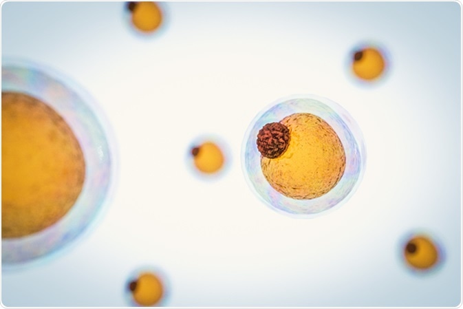
3d render of fat cells. Image Credit: CI Photos / Shutterstock
So far, the negative effects of white fat accumulation have been well studied, but it remains unclear how beige and brown adipocytes interact with their environment to modulate energy use.
There is renewed and growing focus on adipose tissue biology, given the role it plays in obesity, which in turn is associated with an increased risk of type 2 diabetes, atherosclerosis, heart disease, cancer and infertility.
A limitation of traditional histological methods is that they provide a superficial glimpse into the environment of adipocytes and do not yield a more in-depth understanding of their functioning – both self and with other cells. The recently developed Adipo-Clear test sheds more light on adipose tissue biology and promises to grow our understanding of fat cells, particularly beige adipocytes.
Adipo-Clear is a new adipose tissue processing technique that allows for 3D visualization of fat cells under fluorescence microscopy. Here are some of the key applications of Adipo-Clear.
Better Understanding of Beige Adipocyte Development
Little is known or understood about the developmental origins of beige adipocytes. The Adipo-Clear test may just be the breakthrough that researchers were so far hoping for. Its use has shown variation in adipocyte tissue organization, which could eventually be used as a tool to gain more insight into the developmental origins of these cells.
Confirming Links Between Fat Cells and the Nervous System
Adipose tissue, in addition to comprising of adipose cells, is also home to certain neuronal processes and innervations that are crucial for thermogenesis. With Adipo-Clear, researchers were able to demonstrate a clear relationship between sympathetic projections in beige adipocytes. This link opens new avenues for research that could explain what mediates this relationship and its mechanism of action. Researchers believe that by selectively activating sympathetic signals to fat, they might be able to successfully turn on these thermogenic cells. This in turn could yield benefits linked to optimal energy use and weight regulation.
New Evidence of the Role of PRDM16 in Metabolism
PRDM16 (PR domain containing 16) is a protein encoded by the PRDM16 gene and acts as a transcription coregulator, controlling the development of brown adipocytes in brown adipose tissue. According to the new Adipo-Clear test, the density of sympathetic projections is clearly dependent on PRDM16 in adipocytes. This dependent relationship was confirmed when PRDM-16 knockout mice were unable to activate beige fat on exposure to thermal stressors, making them susceptible to metabolic disease. This finding indicates another potential mechanism behind the many metabolic benefits mediated by PRDM16.
In conclusion, over the last two decades, researchers have identified numerous genes and pathways involved in the regulation of brown and beige adipocyte biology. Each discovery has provided deeper understanding of fat cell biology and indicated several promising therapeutic targets for metabolic diseases. With the growing incidence of obesity and its associated metabolic conditions, both in the developed and developing world, tests such as Adipo-Clear offer hope to yield further insight in this field and enable researchers to devise successful targets for non-communicable disease therapy.
Sources
- Mochizuki N, Shimizu S, Nagasawa T, Tanaka H, Taniwaki M, Yokota J, Morishita K (November 2000). "A novel gene, MEL1, mapped to 1p36.3 is highly homologous to the MDS1/EVI1 gene and is transcriptionally activated in t(1;3)(p36;q21)-positive leukemia cells". Blood. 96 (9): 3209–14.
- Three-Dimensional Adipose Tissue Imaging Reveals Regional Variation in Beige Fat Biogenesis and PRDM16-Dependent Sympathetic Neurite Density. Jingyi Chi et al. Cell Metabolism. Volume 27, Issue 1, p226–236.e3, 9 January 2018, http://www.cell.com/cell-metabolism/abstract/S1550-4131(17)30724-6
- Brown and beige fat: development, function and therapeutic potential. Matthew Harms & Patrick Seale. Nature Medicine volume 19, pages 1252–1263 (2013). Retrieved from https://www.nature.com/articles/nm.3361
- Fat in 3D. Danielle Gerhard. Biotechniques. The International Journal of Life Science Methods. https://www.future-science.com/btn/news/feb18/21
- The significance of beige and brown fat in humans. Florian W Kiefer. Endocr Connect. R70–R79. (2017). https://www.ncbi.nlm.nih.gov/pubmed/28465400
Last Updated: Feb 26, 2019