A team from the University of Toronto have pioneered a new of rapidly sizing and counting cells. Strohm et al. combined a microfluidics-based single-cell stream with ultrasound acoustics to yield measurable ultrasound echoes of single cells.
Their approach uniquely compares these distinct spectral features with established theoretical models to achieve a means of accurate cell sizing. The trailblazing method relies on multi-parameter cellular characterization using fluorescence, light scattering, and quantitative photoacoustic techniques.
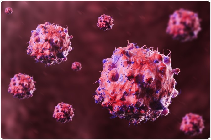
 ImageFlow | Shutterstock
Microfluidics magic
Since the invention of flow cytometry in the 1960s, characterization of cells has enabled high-throughput characterization of cells which have revolutionized several research fields. Microfluidics-based technology has supplemented flow cytometry since - owing to its simplicity, small device size, and integration with a range of instrumentation and analytical tools.
Microfluidic-based cell counters and sorters classify cells via several approaches; the most conventional using light scattering to the lesser-known that use acoustic waves. Of them, electrical impedance (the resistance to a current generated when voltage is applied) is the only known determiner of absolute cell size. The gold standard method driven by this approach is the Coulter Counter.
Other approaches also suffer limitations, for example, FACS only determines the relative sizes of cells in a systematic manner, which limits resolution. Similarly, other systems that use dynamic light scattering, laser diffraction, or bulk acoustic scattering approaches are also relative measures of cell size.
These techniques require prior knowledge of optical and/or acoustic sample properties and require bulk sample approximation which eliminates individual cell size measurements from being collected.
Flow cytometry systems are also limited by inertial, electrokinetic, acoustophoretics and surface acoustic waves used to sort cells by size and/or density differences.
For these reasons, a non-invasive approach to single-cell counting and sizing that affords the simplicity of a microfluidic system is highly sought-after.
The ultra-approach to absolute cell sizing
Ultrasound approaches have been used historically to quantify tissue properties and aid in the diagnosis of diseases. The inference of bulk-tissue properties is achieved at low frequency, however; higher frequencies are required to probe single cells.
The methodology behind modeling the scattering behavior of spherical objects is well developed. Using this scattering theory, Strohm and colleagues previously demonstrated that sizing of micro-sized particles can be achieved using pulse-echo ultrasound. However, cells evaded the ability to be characterized owing to the low frequencies at which the system operated.
Now, a paper published in Nature Scientific reports outlines high-throughput microfluidic-based acoustic flow cytometer capable of sizing single cells. The team custom-designed a polydimethylsiloxane PDMS device flow which hydrodynamically focusses cells into a narrow stream (10 × 10 μm) in both the lateral and axial directions.
As the cells moved single file under the ultrasound transducer operating at 375MHz, ultrasound echo from each passing cell was recorded by custom ultrasound hardware and software. The technique combines a narrow stream with fast ultrasound pulsing and acquisition with sub-nanosecond resolution.
The team validated the system using a solution of polystyrene microspheres. The ultrasound signal measured from a single 3 μm polystyrene microsphere was in excellent agreement with the theoretical spectrum indicating accurate validation of cell measurements for cells. The team used 2 cell lines: (1) acute myeloid leukemia (AML) cells, and (2) HT29 colorectal cancer cells.
Each demonstrated the validity for cell measurements of varying sizes. A single high-frequency ultrasound signal from each cell type possessed distinct minima at specific frequencies that are related to the cell size. The AML and HT29 cells measurements were calculated using a peak detector algorithm measured 10.0 ± 1.7 μm and 15.0 ± 2.3 μm, respectively. These measurements were in good agreement with those predicted using electrical impedance, validating the role of acoustic flow cytometry as an additional method of determining the absolute size of cells on a cell by cell basis.
This proof-of-concept study demonstrates that ultra-high frequency ultrasound can be integrated into a microfluidic device to achieve absolute single-cell sizing. Throughput improvements can be made by way of increasing flow speed, the frequency of ultrasound repetition and aby applying parallel signal recording and analysis techniques for continuous operation.
The ultrasound hardware can be integrated into a wide variety of other microfluidic-based cell characterization systems, potentially enabling a multiparameter analysis of the cells to extract additional cellular details to further enhance cell classification."
As of yet, cell morphology cannot be ascertained by flow cytometry alone; by implementing a laser, photoacoustic measurements can be taken simultaneously with ultrasound paving the way for several telling parameters to be measured.
These range from cell quantification to investigation of the morphological, functional and mechanical properties of cells via label-free or labeled (targeted nanoparticles, fluorescent and colorimetric dyes) interrogation of cell properties.
These include the calculation of the nucleus-to-cytoplasmic ratio of cells in a label-free manner, blood counts and red blood cell morphology quantification. The of the team’s work bodes exciting prospects in the future as it affords an added versatility that the gold standard Coulter Counter cannot.
Source
Strohm et al. (2019) Sizing biological cells using a microfluidic acoustic flow cytometer. Scientific Reports. https://doi.org/10.1038/s41598-019-40895-x
Further Reading
Last Updated: Jul 18, 2019