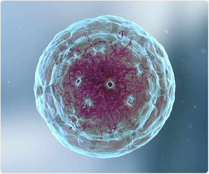
Autophagy is the name given to the mechanism by which cells destroy their own cellular components, often for the purpose of recycling the material and replacing damaged organelles. Commonly degraded components include aggregated proteins and ribosomes, but also other cellular constituents.
 YuriiHrb | Shutterstock
Three primary types of autophagy are macro and micro-autophagy, as well as chaperone-mediated autophagy. Macro-autophagy moves the cytoplasmic component to its lysosome using a double-membrane vesicle, called a phagophore, generated by the endoplasmic reticulum.
On the other hand, micro-autophagy does not use a separate vesicle and instead takes up cellular components directly into the lysosome. Finally, chaperone-mediated autophagy utilizes specialized carrier proteins that are recognized by proteins on the outer membrane surface of the lysosome to transport proteins selected for degradation into the compartment.
Particular proteins either promote or inhibit autophagy within cells. UVRAG, BIF-1, and Atg14L are examples of autophagy promoters, while Rubicon and Bcl-2 are autophagy inhibitors.
Acid proteases present in the lysosome break down the targets of autophagy to their constituent amino acids and other products that are transported to the cytoplasm by using transporter proteins following degradation, before being re-used to assemble new proteins and organelles.
Why does autophagy occur?
Cellular components may become damaged over time, necessitating their replacement. Damaged organelles may not only lose their funtion, but may also become hyper or hypo-active, leading to a cascading series of damaging effects.
Autophagy has been suggested as a natural anti-cancer mechanism, as it limits necrosis and inflammation, and prevents cells with defective components or DNA from replicating. However, in some cases, it has been shown that autophagy may act to promote cancer cell adaptation and even promote drug resistance.
It should be noted that since autophagy is a natural cellular process, it is capable of defective function as with any biomechanical operation. For example, many cancers exhibit deletion of autophagy promotor protein UVRAG, leading to fewer autophagy events taking place within the cell.
Other examples of material that is destroyed via autophagy include proteins present in excess, proteins that have become denatured or have aggregated, as well as invasive bacteria and viruses.
Autophagy is not only important for the removal of improperly functioning cellular components, but is also essential to provide materials with which the cell is fueled during periods of protracted starvation.
What triggers autophagy?
The most common initiating factor for autophagy (especially in non-mammalian cells) is a lack of nutrients, meaning that the recycling of cell components is a critical source of material for cell function. Yeasts in particular have been studied extensively for autophagy triggers, and a lack of nitrogen, glucose, amino acids, and zinc have been shown to initiate non-selective autophagy.
In mammalian cells, lack of only some amino acids appears to initiate autophagy in cultures, though this is also heavily dependent on the type of cell and tissue source. The endocrine system is thought to organize autophagy on the larger scale of the whole organism. Insulin, for example, suppresses autophagy in the liver, while glucagon encourages it.
Other sources of stress to the cell, such as the presence of quickly replicating microorganisms, can also initiate autophagy. Various types of bacteria, including Streptococcus pyogenes, have been shown to be targets of macro-autophagy within cells.
Further Reading
Last Updated: Mar 14, 2019