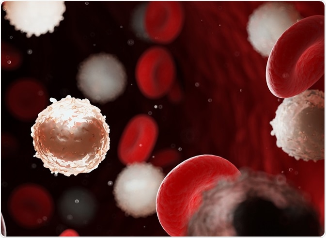
Researchers have developed a new methodology for the characterization of circulating leukemia cells, and other low-abundance cells, by flow cytometry. The technique enables quantification of fluorescence signatures that provide the basis of immunophenotyping - the identification of cells or cell subsets by their cell-surface or intracellular marker profile.
Termed time-delayed integration–spectral flow cytometry (TDI-SFC), the method combines the high resolution of spectral flow cytometry (SFC) with a CCD camera operating in TDI mode.
This article will cover:
 Sebastian Kaulitzki | Shutterstock
Limitations of multiparameter flow cytometry
Multiparameter flow cytometry allows several cell features which include granularity, protein expression and size, to characterize cells. Each of these features is tracked by means of fluorescence labelling, followed by spectral sorting hence the term ‘multiparametric’.
Multiparameter flow cytometry cell sorting is achieved by ‘gating’, separating cells based on scatter as well as markers delineated by fluorescence labeling. This sorting ability is contingent on cell number, requiring >10 000 cells to establish the correct thresholds as well as prevent crosstalk during spectral sorting. In the absence of sufficient cell numbers, poor spectral resolution results and ‘noise’ generated from unusual autofluorescence events can result in cell misclassification.
Fluorescence microscopy circumvents misclassification as it uses morphologically- localized fluorescence. However, cells are counted manually, which severely limits the clinical throughput for samples exceeding a total of 100 cells. Although Imaging flow cytometers (IFCs) solve this problem they are expensive and data analysis is semi-automatic, still limiting clinical processing.
Introducing time-delayed integration–spectral flow cytometry (TDI-SFC)
A solution to this unique problem has been presented by Hu et al comes in the form of SFC; this version of flow cytometry enables the analysis of 100–10 000 cells/sample. In combination with TDI, the proportion of time during which the system operates (duty cycle) and the sensitivity are both improved.
CCD cameras convert emission photons into photo charges which comprise an array of pixels. In TDI, the photocharges are shifted downwards from one pixel to another, in synchrony with the direction and velocity of the cell in the flow, allowing the photocharge signal to spread over many pixels.
This effect is analogous to camera panning and prevents image blur and is referred to as signal integration. The use of fluorescent beads optimizes the synchronization of TDI with cell velocity; overall this provides three distinct advantages
- The signal-to-noise ratio is improved by a factor of N when the signal is integrated over N rows compared to standard full-frame readouts.
- When cells exit the CCD camera field, the integrated emission spectrum is sent for readout; in this way the shutter is never closed. This is particularly problematic when the total available cell count is limited by the number in the sample (<10 000) as cells can escape CCD detection.
- Multiple cells can be resolved.
Harnessing the power of TDI-SFC
TDI-SFC is simple and able to combine several signals in a what is referred to as ‘multiplexing’ and it lends its self well to the clinical sampling of high-volume, low-abundance cells such as circulating leukemia cells and circulating tumor cells. The researchers presented circulating leukemia cell immunotyping as a proof-of-concept application.
Circulating leukemia cell immunotyping is especially relevant in cases of patients in remission – circulating leukemia cells may develop drug resistance, which can evolve into lethal levels of measurable residual disease.
Traditional fluorescence microscopy and multiparameter flow cytometry-based analysis has previously failed to succeed due to (i) background noise originating from blood cells and (ii) the low abundance of circulating leukemia cells. TDI-MFC can include a flow-sorter to collect immunonotyped circulating leukemia cells which prevents laser-dissection or single-cell picking for circulating leukemia cell recovery necessary when sorting them via microscopy.
Hu et al. demonstrated the utility of TD-SFC for a B-ALL cell line stained with a nuclear dye and a leukemic marker (anti-TdT-FITC). 100% of cells were classified accurately by reading spectra with high signals. The signal-to-noise ratio generated by TDI alone was deconvoluted by SFC resulting in identification even at low cell numbers (46–111 cells). The fluorescence sensitivity was improved by the excitation intensity, epi-illumination numerical aperture, and integration time.
A spanner in the works?
The authors note some limitations with this TDI-system, however. The throughput is limited by the readout rate of the CCD and the coefficient of variation which measures precision and repeatability significantly higher (27.8%) than that achieved by multiparameter flow cytometry (6.1%).
The authors speculate that the use of 2D rather than 1D microfluidic focusing will allow cells to be reproducibly focused to align the cells laterally and subsequently lower the coefficient of variation.
The authors hope that following further optimization of TDI-SFC, it could potentially be used ‘in any application requiring the immunophenotyping of low-abundance cells, such as in monitoring measurable residual disease in acute leukemias following affinity enrichment of circulating leukemia cells from peripheral blood’.
Source:
Hu et al. (2019) Time-Delayed Integration–Spectral Flow Cytometer (TDI-SFC) for Low-Abundance-Cell Immunophenotyping. Analytical Chemistry. https://doi.org/10.1021/acs.analchem.9b00021
Further Reading
Last Updated: Jul 26, 2019