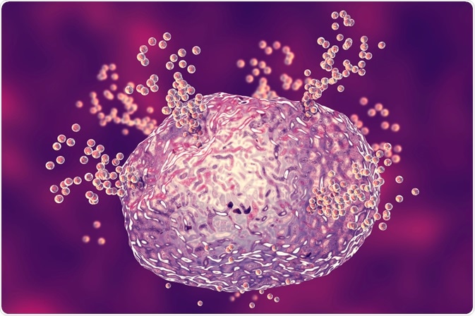
Flow cytometry is a technique used by biologists to observe populations of cells with high precision and accuracy.
 Kateryna Kon | Shutterstock
Flow cytometry works by illuminating cells, or other types of particle, as they flow in front of a light source, such as a single or dual laser beam. The light source then detects and correlates signals from those cells. By combining data acquisition speed, sample dimension, precision, and measurement multiplicity, flow cytometry physically separates sub-populations of groups of cells.
How is flow cytometry used in immunology?
Single-cell responses
In immunology, this technique can be used to generate detailed profiles of single-cell responses to different stimuli (e.g. phosphoproteins) in T-cells, B-cells, myeloid cells, and several clinically relevant cytokines, such as GM-CSF, IFN-γ, IL-10, IL-2, and lipopolysaccharides (LPS) at key regulatory interfaces (e.g. MAPK pathways).
Diagnosing primary immunodeficiencies
Flow cytometry can be used to study specific cell populations and sub-populations, as well as intracellular proteins, cell surfaces, certain functional immune characteristics, and biological effects associated with particular immune defects. Each of these factors can be used to diagnose primary immunodeficiencies.
Immune competence
In a recent study, phagocytosis by leukocytes in whole blood was hypothesized as a potential trait of immune competence in chickens. A whole blood phagocytosis assay using flow-cytometry techniques was performed and assessed using blood from chickens with all four different major histocompatibility complex haplotypes: B21, B19, B15, and B12.
The study used fluorescent latex beads alongside two different serotypes of fluorescently-labeled bacteria that had been killed using heat: Salmonella typhimurium and Salmonella infantis.
The assessment of the phagocytic activity in the whole blood was completed using a no-wash, no-lyse flow cytometry. The results showed that both of the bacteria serotypes were phagocytosed effectively by leukocytes in the blood cultures, whereas the latex beads were not affected.
Why is flow cytometry used in immunology?
Flow cytometry has been conventionally associated with the use of monoclonal antibodies to identify immuno-competent cells, to quantify changes in expression of surface determinants, and to separate cell population subsets before testing their functional characteristics.
Single and dual-laser systems, developments of multicolor fluorescence, and improvements of computing systems for multi-parametric analysis helps in the improvement of diagnosis, prognosis, and monitoring therapies given. Advances in flow cytometry now allow for automated, multiparametric analyses of thousands of samples in a day.
Each of these data sets consist of extremely detailed, multidimensional descriptions of millions of individual cells. The ability to collect this data is currently outpacing the means for data handling and analysis by computers. However, along with improving computer speeds and storage capabilities, flow cytometry could facilitate the field of immunological research.
Further Reading
Last Updated: Feb 14, 2019